Copyright
©2009 The WJG Press and Baishideng.
World J Gastroenterol. Jun 14, 2009; 15(22): 2794-2799
Published online Jun 14, 2009. doi: 10.3748/wjg.15.2794
Published online Jun 14, 2009. doi: 10.3748/wjg.15.2794
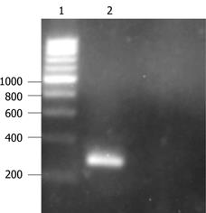
Figure 1 Identification of Ad-p27mt by PCR.
1: 200bp marker; 2: Ad-p27mt.
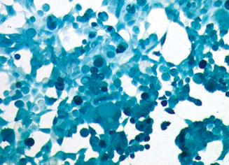
Figure 2 X-gal chemical staining detected the infection efficiency of Ad-p27mt (MOI 50).
Figure 3 Western blotting of expressed p27 protein.
1: Non-infected Lovo cells; 2: Ad-lacZ infected Lovo cells; 3: Ad-p27mt infected Lovo cells.
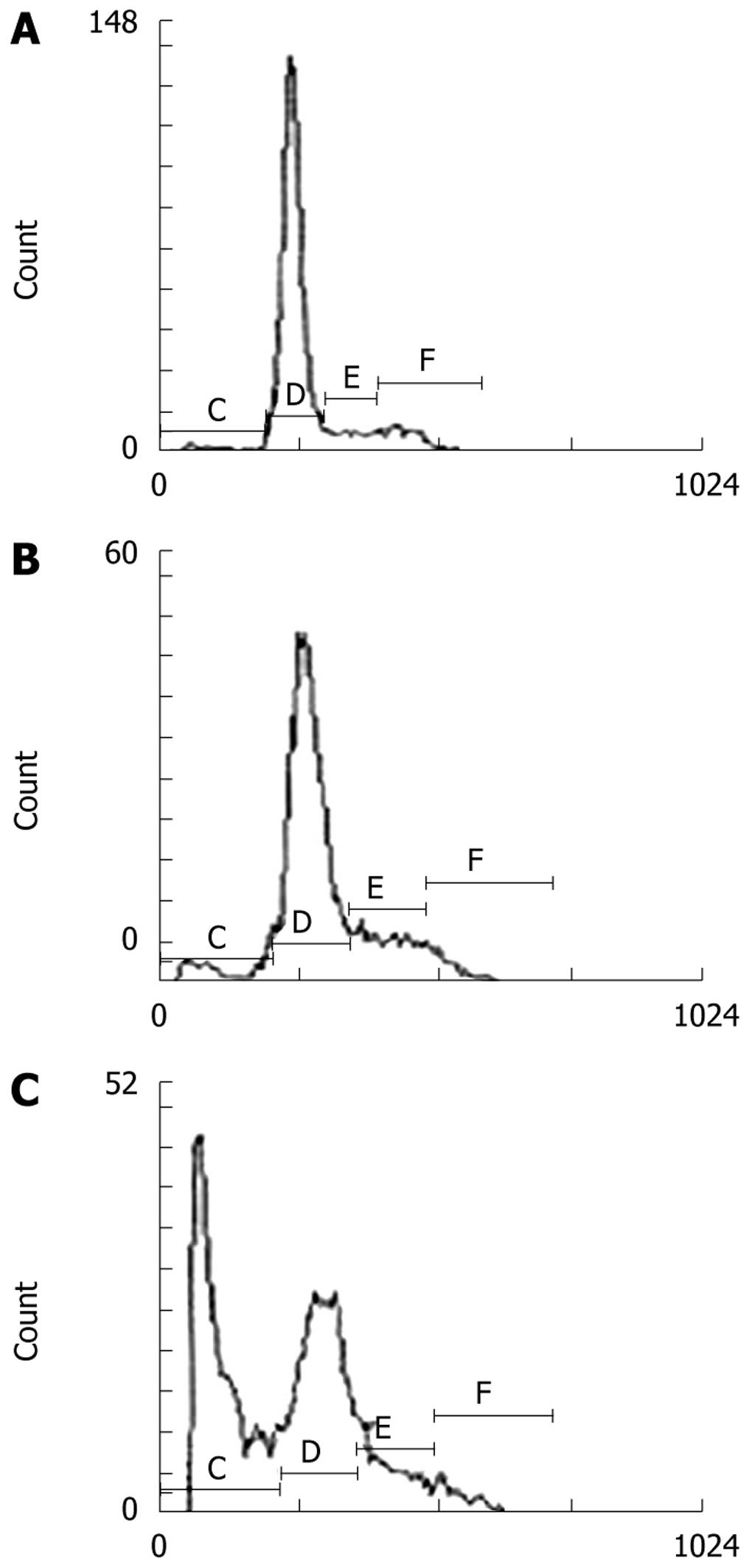
Figure 4 Determination of cell apoptosis by Flow cytometry.
A: Non-infected Lovo cells; B: Ad-lacZ infected Lovo cells; C: Ad-p27mt infected Lovo cells.
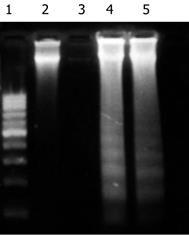
Figure 5 Determination of cell apoptosis by DNA fragment analysis.
1: Marker; 2: Blank group; 3: Ad-lacZ group; 4, 5: Ad-p27mt group.
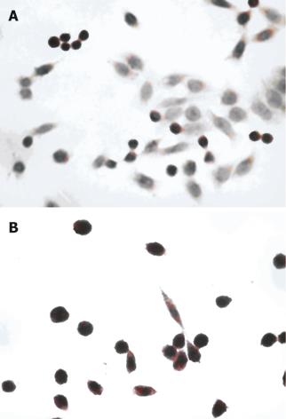
Figure 6 Detection of cell apoptosis by the TUNEL method.
A: Blank group; B: Ad-p27mt.
-
Citation: Chen J, Ding WH, Xu SY, Wang JN, Huang YZ, Deng CS. Effect of
p27mt gene on apoptosis of the colorectal cancer cell line Lovo. World J Gastroenterol 2009; 15(22): 2794-2799 - URL: https://www.wjgnet.com/1007-9327/full/v15/i22/2794.htm
- DOI: https://dx.doi.org/10.3748/wjg.15.2794


