Copyright
©2009 The WJG Press and Baishideng.
World J Gastroenterol. Jun 14, 2009; 15(22): 2754-2762
Published online Jun 14, 2009. doi: 10.3748/wjg.15.2754
Published online Jun 14, 2009. doi: 10.3748/wjg.15.2754
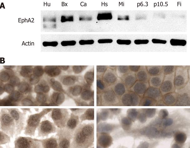
Figure 1 EphA-2 expression in human cell lines.
A: Analysis of EphA2R expression by Western blotting in human umbilical vein endothelial cells (Hu), human pancreatic cancer cell lines BxPc-3 (Bx), Hs766-T (Hs), Capan-1 (Ca), MIA PaCa-2 (Mia), p6.3, p10.5 and human fibroblasts (Fi). EphA2R was detected using a monoclonal antibody and detection of actin levels was performed as a loading control; B: Immunolocalization of EphA2R in human pancreatic cancer cell lines BxPc-3 (left top), Capan-1 (right top), Hs766-T (bottom left), MiaPaca-2 (bottom right). Cells were fixated with methanol, acetone, and water, and a directed monoclonal antibody was used to detect EphA2R using a goat anti-mouse labeled with PO to perform DAB detection. Magnification (× 600), except for MiaPaca-2 (× 400).
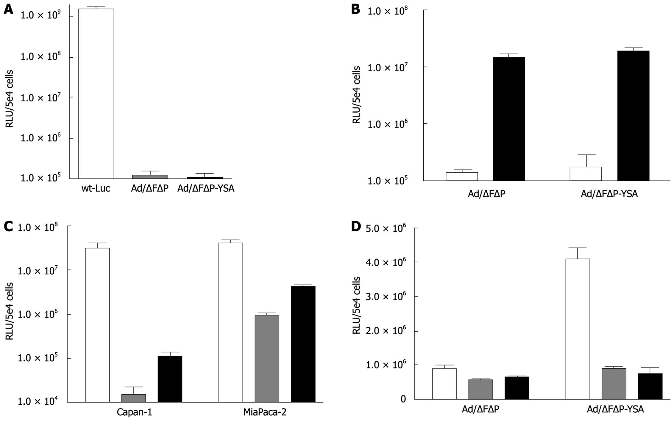
Figure 2 YSA redirects adenovirus to the EphA2 receptor.
A: Transduction of CAR-expressing A549 cells with wild-type, ablated and YSA-retargeted adenoviral vectors with 1000 gc/cell: wt-Ad-Luc, Ad-Luc/∆F(FG)∆P and Ad-Luc/∆F(FG)∆P-YSA. Twenty four hours after infection cells were lysed to determine luciferase levels. Data are expressed as mean ± SD (n = 3). B: Incubation with bi-specific antibody restores infectivity of ablated adenoviral vectors in human fibroblasts. Cells were transduced with 500 gc/cell of Ad-/∆F(FG)∆P or Ad-/∆F(FG)∆P-YSA with (black bars) or without (white bars) EGF receptor targeted bi-specific scFV molecules. Luciferase activity was measured after 24 h and data are expressed as mean ± SD (n = 3). C: Insertion of YSA peptide in the HI loop of ablated adenoviral vector partially restores transduction of human pancreatic cancer cell lines Capan-1 and MiaPaca-2. Cells were transduced with 1000 gc/cell of wt-Ad-Luc (white bars), or Ad-Luc/∆F(FG)∆P (gray bars) or Ad-Luc/∆F(FG)∆P-YSA (black bars). Luciferase activity was measured after 24 h. Data are expressed as mean ± SD (n = 3). D: Pre-incubation with synthetic peptide blocks YSA-mediated targeting of human pancreatic cancer cell line MiaPaca-2. Cells were preincubated with 250 (grey bars) or 500 (black bars) &mgr;mol/L synthetic YSA peptide and transduced with 500 gc/cell of Ad-Luc/∆F(FG)∆P or Ad-Luc/∆F(FG)∆P-YSA. Luciferase was measured after 24 h. Data are expressed as mean ± SD (n = 3).
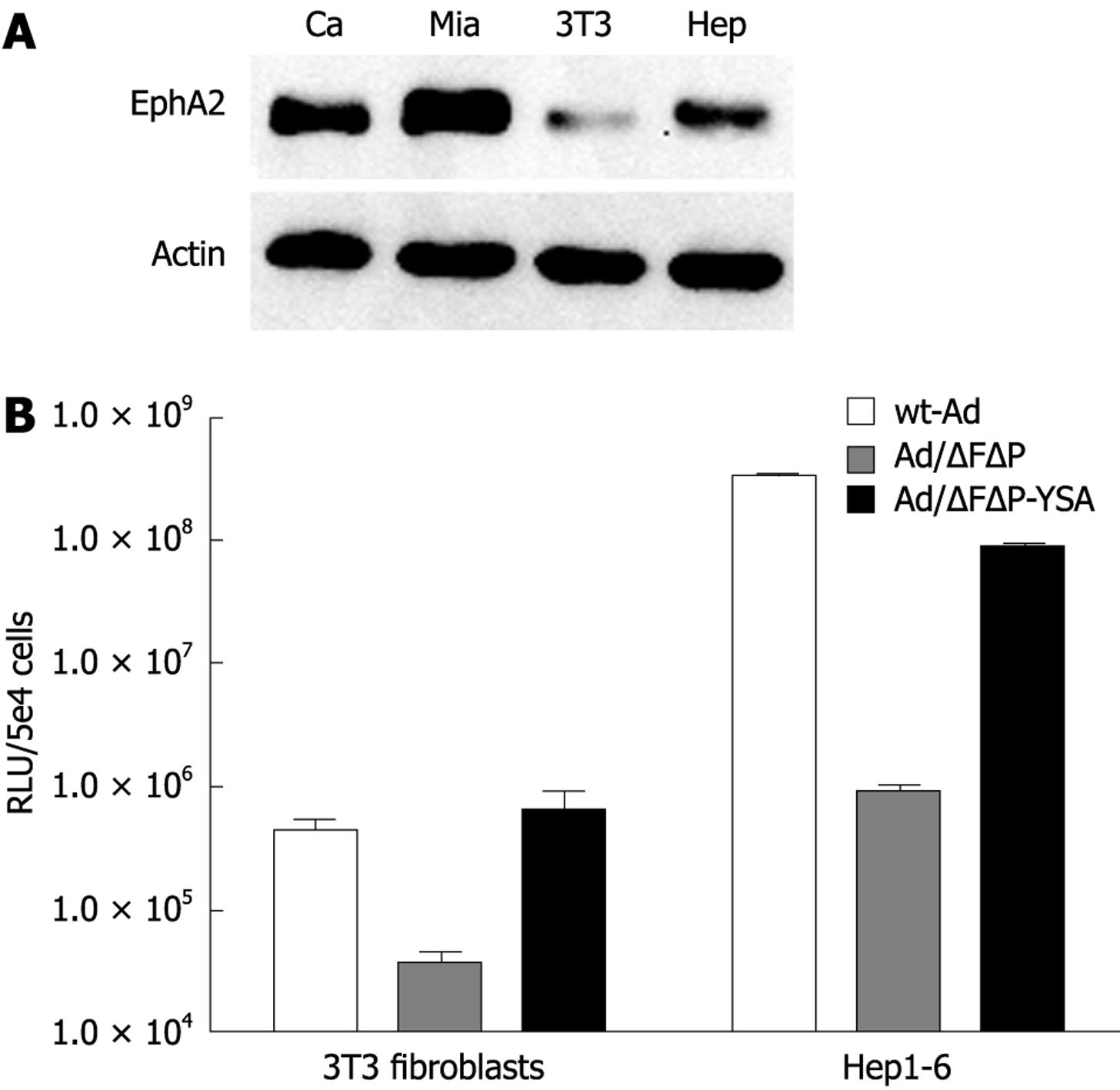
Figure 3 YSA targets adenovirus to cells expressing murine EphA2R.
A: Western blotting demonstrates that mouse 3T3 and Hep1-6 cells, like human pancreatic cancer cell lines Capan-1 and MIA PaCa-2, express the EphA2R. EphA2R was detected using a monoclonal antibody and the housekeeping protein actin was used as a loading control. B: Efficient transduction of mouse 3T3 and Hep1-6 cells by YSA-retargeted adenoviral vector. Cells were transduced with 1000 gc/cell of wt-Ad-Luc (white bars), Ad-Luc/∆F(FG)∆P (gray bars) or Ad-Luc/∆F(FG)∆P-YSA (black bars). Luciferase was measured after 24 h. Data are expressed as mean ± SD (n = 3).
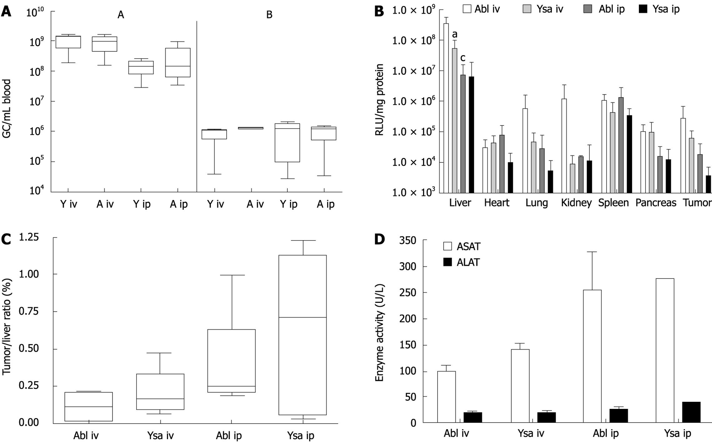
Figure 4 Lack of YSA specific targeting in nu/nu mice.
A: 1 × 10¹¹ gc of Ad/∆F(FG)∆P (ablated, A) or 1 × 10¹¹ gc of Ad/∆F(FG)∆P-YSA (YSA; Y) are rapidly cleared from blood after intravenous (i.v.) and intraperitoneal (i.p.) administration into mice. At 10 min after i.v. or 90 min after i.p. injection and after 3 d (both), adenoviral genomic copies were determined in whole blood with real time PCR. B: Bio-distribution Ad/∆F(FG)∆P and Ad/∆F(FG)∆P-YSA injected i.v. or i.p. into nu/nu mice with a subcutaneous human pancreatic tumor. Animals were sacrificed 3 d after injection of 1 × 10¹¹ gc of adenoviral vector. All organs were harvested and analyzed for luciferase expression/mg of protein. (aP < 0.05 compared with Ad/∆F(FG)∆P after i.v. injection; cP < 0.05 compared with Ad/∆F(FG)∆P after i.v. injection). C: The tumor/liver ratio of luciferase expression in each mouse demonstrates the lack of significant targeting of retargeted adenovirus to pancreatic cancer in vivo. D: Similar ASAT and ALAT levels in serum at 3 d after injection indicates comparable liver toxicity of ablated and retargeted adenoviral vectors in nu/nu mice. Data represent the mean ± SD of 4-7 mice.
-
Citation: van Geer MA, Bakker CT, Koizumi N, Mizuguchi H, Wesseling JG, Oude Elferink RP, Bosma PJ. Ephrin A2 receptor targeting does not increase adenoviral pancreatic cancer transduction
in vivo . World J Gastroenterol 2009; 15(22): 2754-2762 - URL: https://www.wjgnet.com/1007-9327/full/v15/i22/2754.htm
- DOI: https://dx.doi.org/10.3748/wjg.15.2754


