Copyright
©2009The WJG Press and Baishideng.
World J Gastroenterol. Jun 7, 2009; 15(21): 2638-2643
Published online Jun 7, 2009. doi: 10.3748/wjg.15.2638
Published online Jun 7, 2009. doi: 10.3748/wjg.15.2638
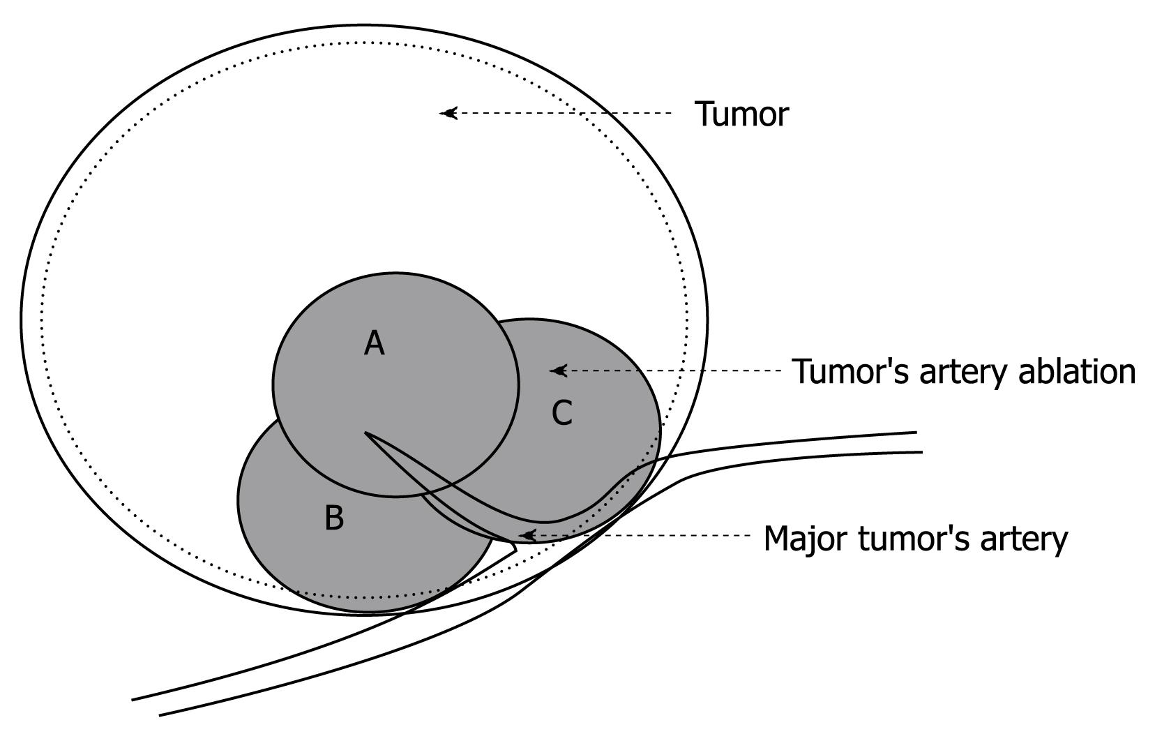
Figure 1 Schematic drawing of PAA using three small ablation foci at the area where the feeding artery entered the tumor, to block the blood supply of HCC.
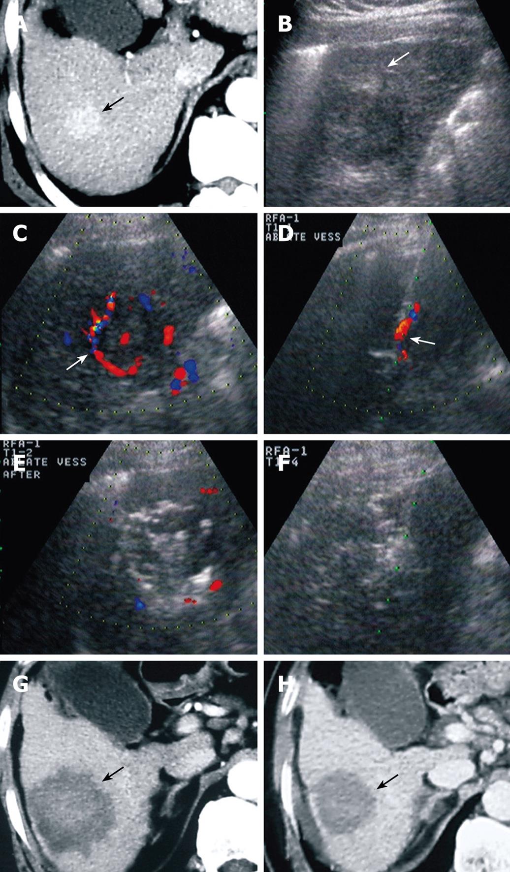
Figure 2 A 65-year-old man with cirrhosis and Child-Pugh class A liver function.
An HCC lesion was diagnosed during regular US examination. A: CT showed a tumor with a size of 3.2 cm × 3.0 cm in the right liver lobe; B: US showed a tumor (arrow) of 4.3 cm × 3.3 cm 3 mo later; C: CDFI showed blood flow into and around the tumor with a velocity of 56.7 cm/s; D: CDFI-guided PAA at the area where the feeding artery entered the tumor (arrow), to block the tumor blood supply; E: After PAA, CDFI showed that the previous feeding artery disappeared and no flow signal within HCC; F: After PAA, RFA was performed in the rest of the tumor; G: Contrast-enhanced CT (1 mo after treatment) showed an ablated area covering the previous tumor, without enhancement; H: Contrast CT (6 mo after treatment) showed no enhancement of the ablated area. The patient has survived more than 10 mo without tumor.
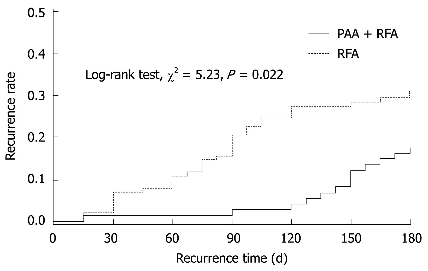
Figure 3 The Kaplan-Meier curves for 6-mo recurrence rate in the two groups.
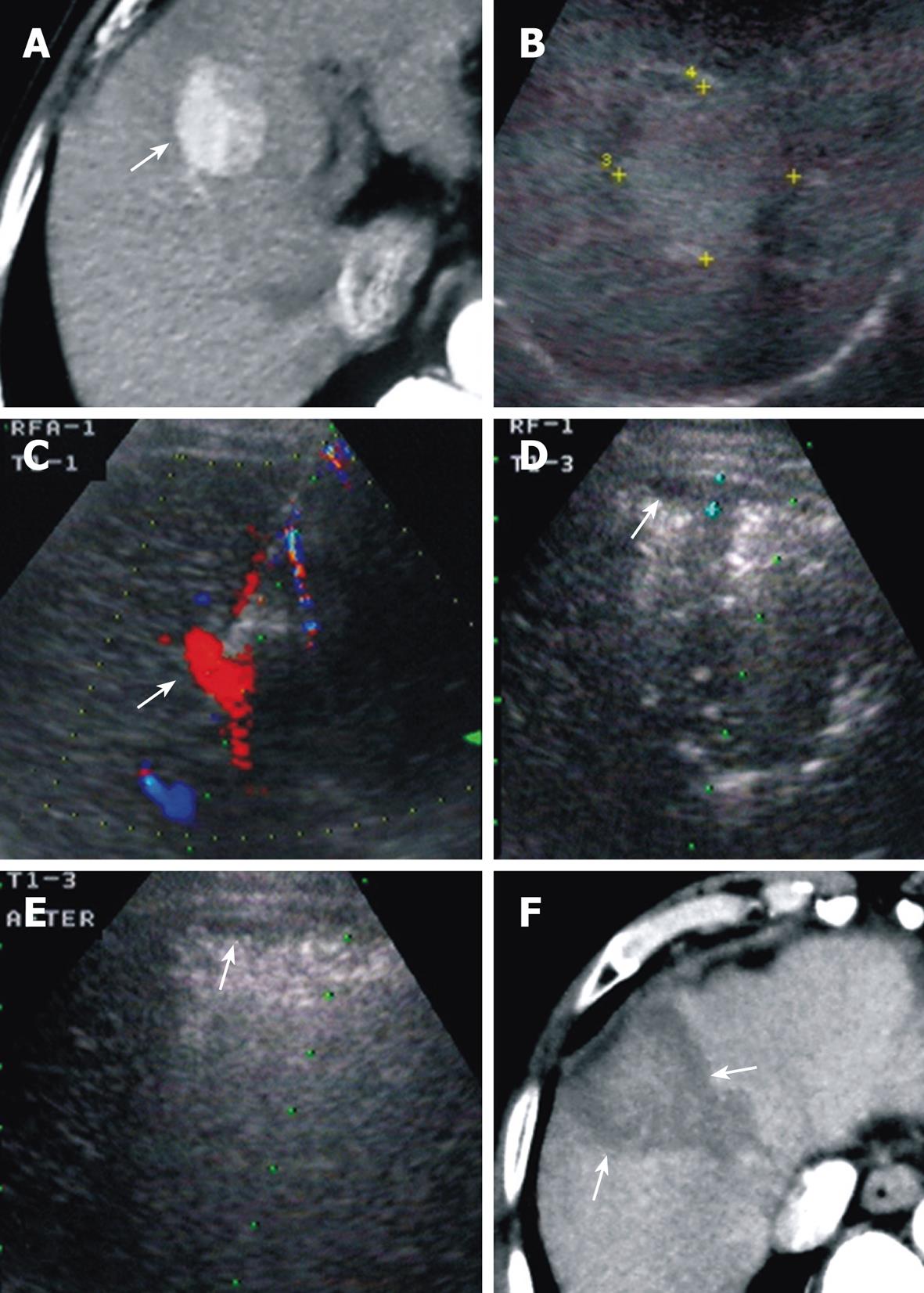
Figure 4 A 72-year-old man with 10 years of hepatitis B was detected with HCC during routine examination.
This patient could not tolerate most of the therapies because of old age and poor general condition. A: Contrast-enhanced CT showed a 3.7 cm × 3.5 cm mass and the feeding vessel (arrow); B: Contrast-enhanced US 2 mo later showed the tumor enlarged to a size of 5.2 cm × 4.2 cm; C: CDFI showed feeding vessels in and around the tumor with a flow velocity of 34.5 cm/s. PAA was performed under CDFI guidance; D: During PAA, a small amount of subcapsular hemorrhage (0.5 cm in depth) was noted; E: The amount of hemorrhage gradually reduced (arrow) after PAA finished; F: Contrast-enhanced CT (24 h after treatment) showed a necrotic area covering the tumor. A small amount of subcapsular hemorrhage was still be noted.
- Citation: Hou YB, Chen MH, Yan K, Wu JY, Yang W. Adjuvant percutaneous radiofrequency ablation of feeding artery of hepatocellular carcinoma before treatment. World J Gastroenterol 2009; 15(21): 2638-2643
- URL: https://www.wjgnet.com/1007-9327/full/v15/i21/2638.htm
- DOI: https://dx.doi.org/10.3748/wjg.15.2638


