Copyright
©2009 The WJG Press and Baishideng.
World J Gastroenterol. Jun 7, 2009; 15(21): 2623-2627
Published online Jun 7, 2009. doi: 10.3748/wjg.15.2623
Published online Jun 7, 2009. doi: 10.3748/wjg.15.2623
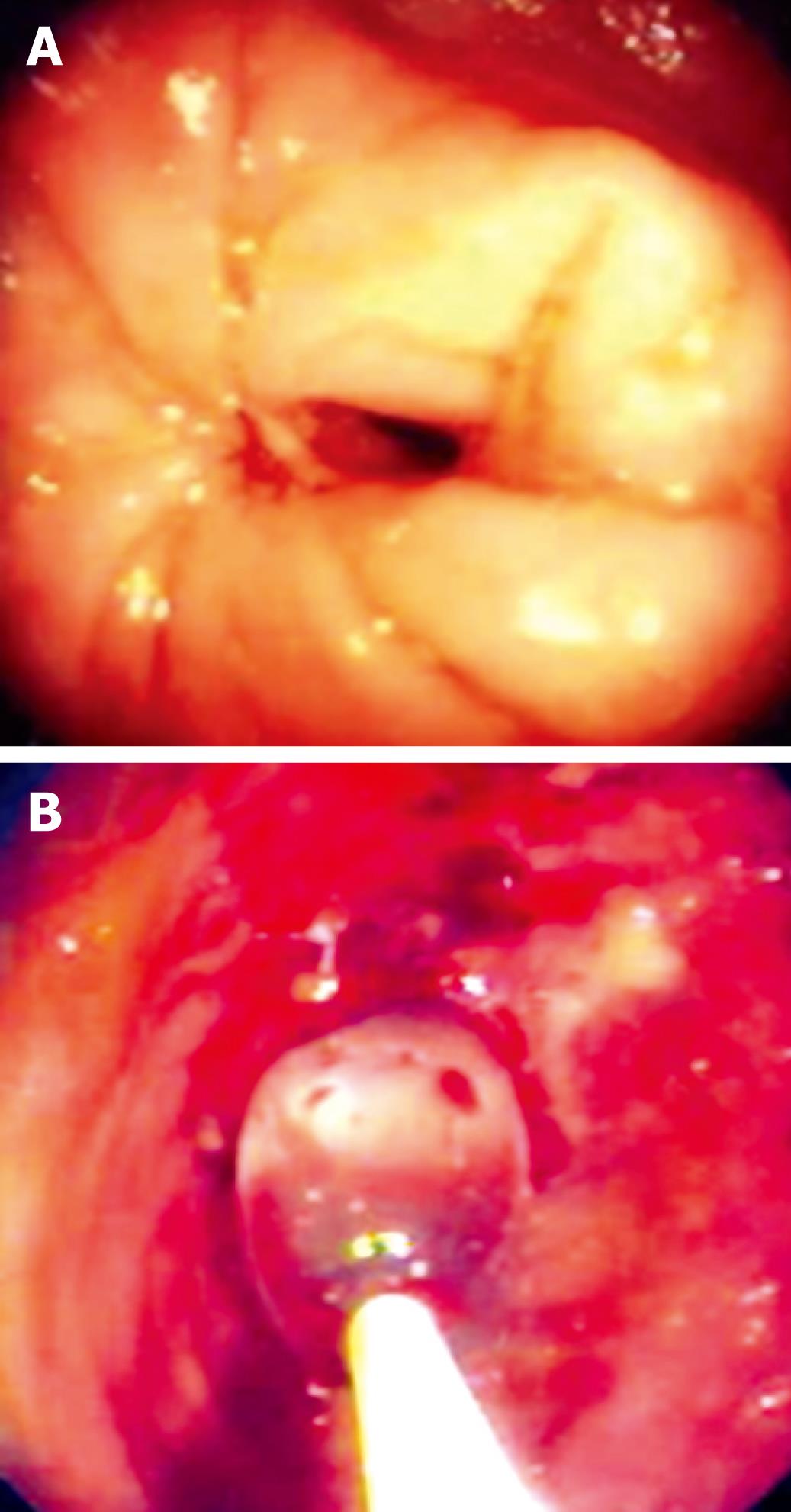
Figure 1 Endoscopic view of a non-passable stricture.
A: Ileum; B: An anastomosis, balloon in the stricture.
Figure 2 Radiological image of the endoscopic dilatation of a short stricture in the ileum.
A: Before dilatation; B: Beginning dilatation; C: Completed dilatation.
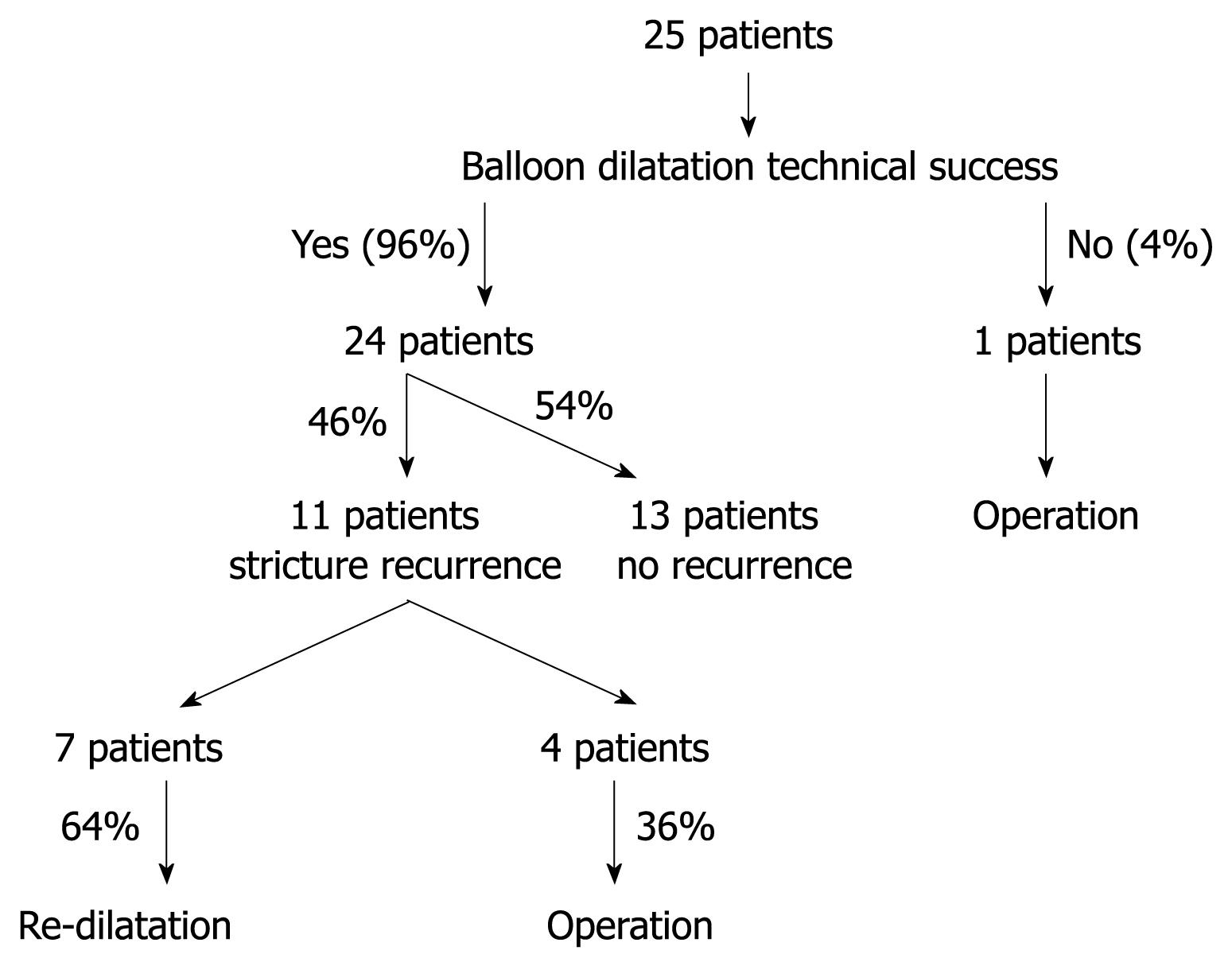
Figure 3 Overview of all patients treated.
- Citation: Stienecker K, Gleichmann D, Neumayer U, Glaser HJ, Tonus C. Long-term results of endoscopic balloon dilatation of lower gastrointestinal tract strictures in Crohn’s disease: A prospective study. World J Gastroenterol 2009; 15(21): 2623-2627
- URL: https://www.wjgnet.com/1007-9327/full/v15/i21/2623.htm
- DOI: https://dx.doi.org/10.3748/wjg.15.2623


