Copyright
©2009 The WJG Press and Baishideng.
World J Gastroenterol. May 28, 2009; 15(20): 2537-2542
Published online May 28, 2009. doi: 10.3748/wjg.15.2537
Published online May 28, 2009. doi: 10.3748/wjg.15.2537
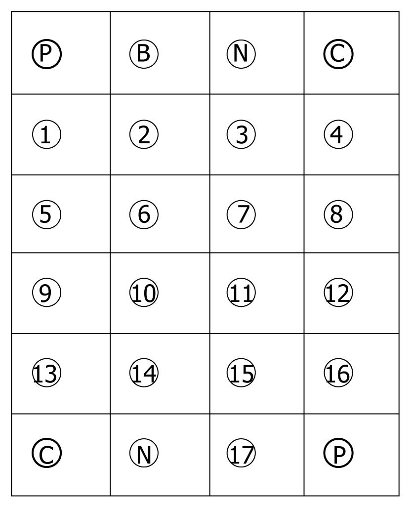
Figure 1 Layout of oligonucleotide probes.
Their sequences are indicated in Table 3.
Figure 2 Partial dual PCR amplification results for intestinal pathogens from fecal samples.
M: DNA marker 2000; B: Blank control.
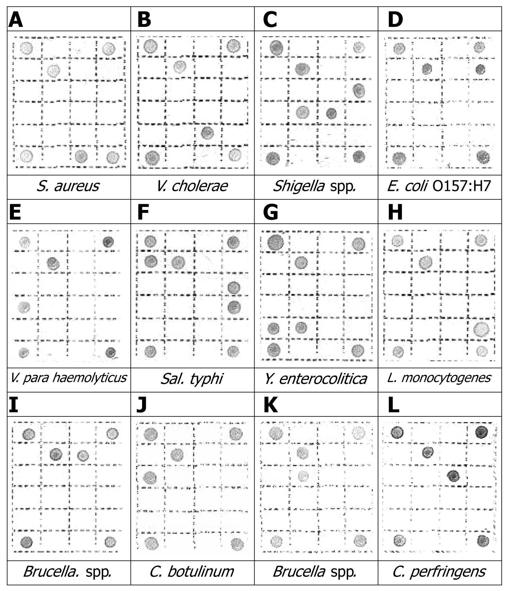
Figure 3 Typical hybridization profiles on the membrane from pure bacterial culture.
- Citation: Xing JM, Zhang S, Du Y, Bi D, Yao LH. Rapid detection of intestinal pathogens in fecal samples by an improved reverse dot blot method. World J Gastroenterol 2009; 15(20): 2537-2542
- URL: https://www.wjgnet.com/1007-9327/full/v15/i20/2537.htm
- DOI: https://dx.doi.org/10.3748/wjg.15.2537


