Copyright
©2009 The WJG Press and Baishideng.
World J Gastroenterol. May 14, 2009; 15(18): 2234-2239
Published online May 14, 2009. doi: 10.3748/wjg.15.2234
Published online May 14, 2009. doi: 10.3748/wjg.15.2234
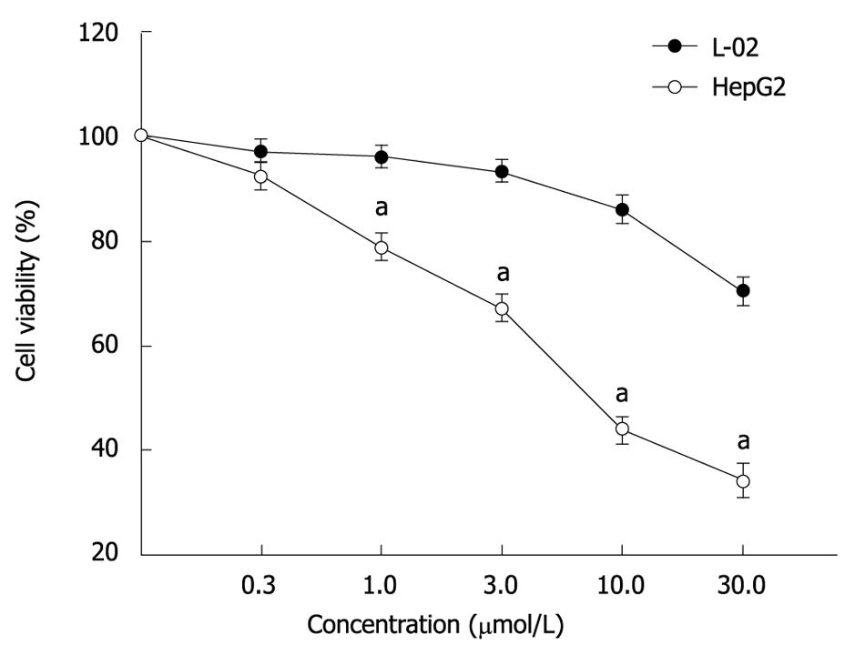
Figure 1 ADFMChR selectively inhibited proliferation of HepG2 cells.
aP < 0.05 vs treatment with ADFMChR in the same concentration to L-02 cells (mean ± SD, n = 9).
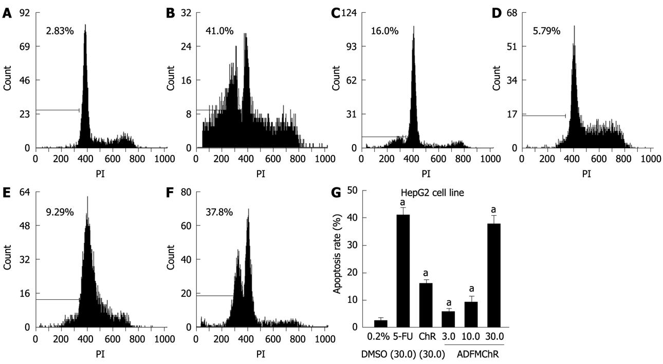
Figure 2 Induction of apoptosis of HepG2 cells by ADFMChR.
A: Treated with 0.2% DMSO; B: Treated with 30.0 &mgr;mol/L 5-FU; C: Treated with 30.0 &mgr;mol/L ChR; D: Treated with 3.0 &mgr;mol/L ADFMChR; E: Treated with 10.0 &mgr;mol/L ADFMChR; F: Treated with 30.0 &mgr;mol/L ADFMChR; G: Quantification of induction of apoptosis analysis of HepG2 cells. aP < 0.05 vs treatment with DMSO (mean ± SD, n = 3).
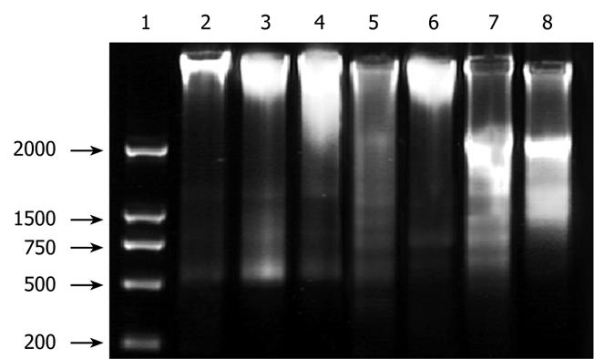
Figure 3 DNA ladder assay showing ADFMChR-induced apoptosis of HepG2 cells.
Lane 1: DNA marker; lane 2: Control; lane 3: 10.0 &mgr;mol/L ADFMChR (24 h); lane 4: 10.0 &mgr;mol/L ADFMChR + GW9662 (24 h); lane 5: 10.0 &mgr;mol/L ADFMChR (48 h); lane 6: 10.0 &mgr;mol/L ADFMChR + GW9662 (48 h); lane 7: 10.0 &mgr;mol/L ADFMChR (72 h); lane 8: 10.0 &mgr;mol/L ADFMChR + GW9662 (72 h).
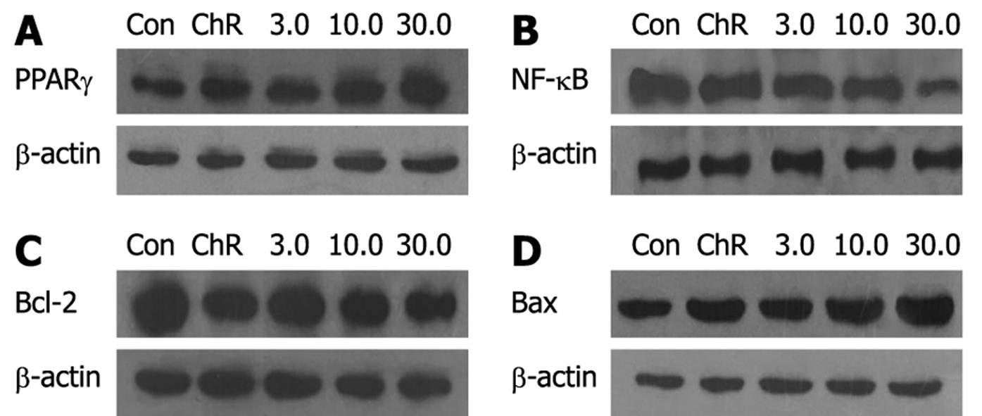
Figure 4 Western blotting analysis showing regulation of PPARγ (A), NF-κB (B), Bcl-2 (C) and Bax (D) protein expression in HepG2 cells by ADFMChR.
(mean ± SD, n = 3).
Figure 5 PPARγ antagonist GW9662 blocked the effects of ADFMChR on PPARγ and NF-κB protein expression in HepG2 cells.
A: PPARγ; B: NF-κB. HepG2 cells were pretreated with 10.0 &mgr;mol/L GW9662 for 30 min, then exposed to 3.0, 30.0 &mgr;mol/L ADFMChR for 24 h, respectively (mean ± SD, n = 3).
- Citation: Tan XW, Xia H, Xu JH, Cao JG. Induction of apoptosis in human liver carcinoma HepG2 cell line by 5-allyl-7-gen-difluoromethylenechrysin. World J Gastroenterol 2009; 15(18): 2234-2239
- URL: https://www.wjgnet.com/1007-9327/full/v15/i18/2234.htm
- DOI: https://dx.doi.org/10.3748/wjg.15.2234


