Copyright
©2009 The WJG Press and Baishideng.
World J Gastroenterol. Apr 21, 2009; 15(15): 1910-1913
Published online Apr 21, 2009. doi: 10.3748/wjg.15.1910
Published online Apr 21, 2009. doi: 10.3748/wjg.15.1910
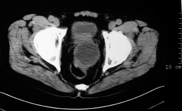
Figure 1 CT scan image at pelvic showing a large low-density lesion arising from the rectum.
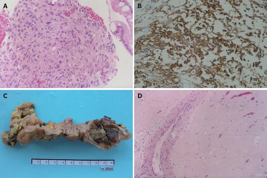
Figure 2 Different images of the rectal tumor.
A: Histopathological microphotograph of biopsy tissue showing spindle-shaped cells with obvious atypia and active mitoses (HE, × 200); B: Envision immunohistochemical stained tumor cells showing strong and diffuse positive staining for CD117 (HE, × 200); C: Macroscopic image of the resected tissue showing a submucosal tumor grayish and uniformly soft tissue texture, measuring 4 cm × 3.5 cm × 3 cm in size; D: Histological examination of the resected tissue showing no residual tumor cells, except for blood vessels and scattered lymphocytes (HE, × 200).
- Citation: Hou YY, Zhou Y, Lu SH, Qi WD, Xu C, Hou J, Tan YS. Imatinib mesylate neoadjuvant treatment for rectal malignant gastrointestinal stromal tumor. World J Gastroenterol 2009; 15(15): 1910-1913
- URL: https://www.wjgnet.com/1007-9327/full/v15/i15/1910.htm
- DOI: https://dx.doi.org/10.3748/wjg.15.1910


