Copyright
©2009 The WJG Press and Baishideng.
World J Gastroenterol. Mar 28, 2009; 15(12): 1524-1527
Published online Mar 28, 2009. doi: 10.3748/wjg.15.1524
Published online Mar 28, 2009. doi: 10.3748/wjg.15.1524
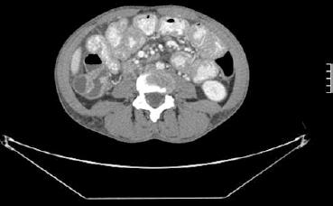
Figure 1 Computerized tomography scan image displaying diffuse lymphadenopathy and small bowel wall thickening.
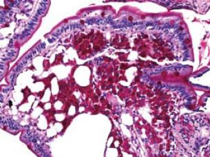
Figure 2 A periodic acid-Schiff (PAS) stain showing extensive PAS positive material within the foamy macrophages (small intestinal biopsy).
- Citation: Karsan SS, Karsan HA, Karsan AS, McMillen JI. A case of Noonan syndrome and Whipple’s disease in the same patient. World J Gastroenterol 2009; 15(12): 1524-1527
- URL: https://www.wjgnet.com/1007-9327/full/v15/i12/1524.htm
- DOI: https://dx.doi.org/10.3748/wjg.15.1524


