Copyright
©2009 The WJG Press and Baishideng.
World J Gastroenterol. Mar 28, 2009; 15(12): 1415-1419
Published online Mar 28, 2009. doi: 10.3748/wjg.15.1415
Published online Mar 28, 2009. doi: 10.3748/wjg.15.1415
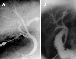
Figure 1 Cystic duct.
A: near a segmental bile duct; B: very close to the sectoral bile duct.
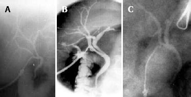
Figure 2 Cystic duct.
A: Cystic duct joining the segmental; B: Sectoral hepatic ducts that could be misinterpreted as cystic ducts; C: Right hepatic ducts that could be misinterpreted as cystic ducts.
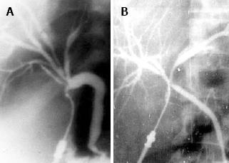
Figure 3 Cystic duct joins the convergence of the sectoral (A) and hepatic ducts (B).
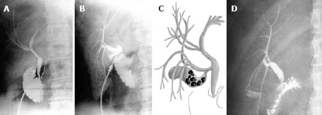
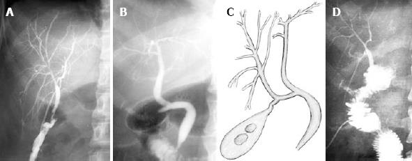
Figure 4 Cholangiography.
A: An operative cholangiography with missing ducts within segments V and VIII; B: Cholangiography of the injured anterior sectoral duct; C: A schematic of the biliary anatomy before the injury took place (the anterior sectoral duct was misinterpreted as a cystic duct); D: Following hepatico-jejunostomy.
Figure 5 Demonstrates a case with an external biliary fistula that required Roux-en-Y repair.
A: Fistulography showing the right hepatic duct; B: ERCP showing the rest of the biliary tree; C: A schematic of the biliary anatomy before the injury took place (the right hepatic duct was misinterpreted as a cystic duct); D: Following hepatico-jejunostomy.
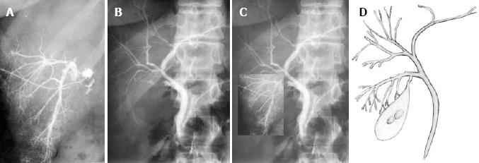
Figure 6 A biliary fistula that resolved spontaneously.
A: Fistulography showing the ducts within segment V; B: ERCP showing the rest of the biliary tree; C: The entire biliary tree if the injury hadn’t take place; D: A schematic of the biliary anatomy prior to injury.
- Citation: Colovic RB. Isolated segmental, sectoral and right hepatic bile duct injuries. World J Gastroenterol 2009; 15(12): 1415-1419
- URL: https://www.wjgnet.com/1007-9327/full/v15/i12/1415.htm
- DOI: https://dx.doi.org/10.3748/wjg.15.1415


