Copyright
©2009 The WJG Press and Baishideng.
World J Gastroenterol. Mar 21, 2009; 15(11): 1401-1403
Published online Mar 21, 2009. doi: 10.3748/wjg.15.1401
Published online Mar 21, 2009. doi: 10.3748/wjg.15.1401
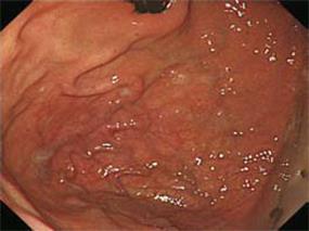
Figure 1 Upper gastrointestinal endoscopy showing a meandering, dilated, pulsatile vessel in the fundus.
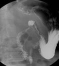
Figure 2 Double contrast view of the duodenum show-ing the acute angulation and stenosis between the duodenal bulb and second portion of the duodenum.
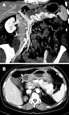
Figure 3 Abdominal CT findings.
A: Oblique sagittal view demonstrates the absence of the splenic vein with confluence of the superior mesenteric vein (arrow) and the presence of the tortuously engorged gastroepiploic vein in the pancreas (asterisk), forming the main portal vein (arrowhead); B: Axial image of the abdomen shows the tortuously engorged gastroepiploic vein in the pancreas.
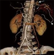
Figure 4 Three dimensional CT image demonstrates the tortuously dilated left gastric artery (arrow) and the left gastroepiploic artery (arrowhead) with non-visualization of the splenic artery.
- Citation: Shin EK, Moon W, Park SJ, Park MI, Kim KJ, Lee JS, Kwon JH. Congenital absence of the splenic artery and splenic vein accompanied with a duodenal ulcer and deformity. World J Gastroenterol 2009; 15(11): 1401-1403
- URL: https://www.wjgnet.com/1007-9327/full/v15/i11/1401.htm
- DOI: https://dx.doi.org/10.3748/wjg.15.1401


