Copyright
©2009 The WJG Press and Baishideng.
World J Gastroenterol. Mar 21, 2009; 15(11): 1398-1400
Published online Mar 21, 2009. doi: 10.3748/wjg.15.1398
Published online Mar 21, 2009. doi: 10.3748/wjg.15.1398
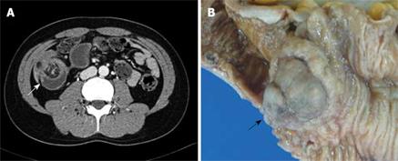
Figure 1 Enhanced abdominal CT and photograph of the pathological specimen.
A: A soft tissue mass with fatty-tissue attenuation in the terminal ileum, and with intussusception is shown (arrow); B: Grossly, an approximate 3 cm × 3 cm × 2.5 cm sized polypoid mass was located in the ileum, 60 cm from the ileocecal valve (arrow).
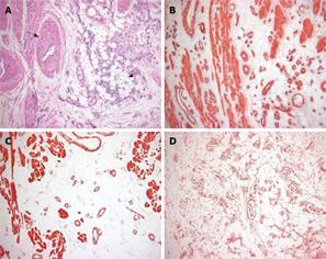
Figure 2 Photomicrograph of the histopathological specimen.
A: The specimen was composed of three tissue components: mature adipose tissue (arrowhead) and smooth muscle surrounding thick-walled, medium-sized, vascular channels (arrow) (HE, × 40); B: Immunohistochemistry (IHC) staining showed that the proliferating smooth muscle cells were positive for α-SMA (IHC, × 40); C: Smooth muscle cells showed immunoreactivity for desmin (IHC, × 40); D: The scattered small blood vessels showed immunoreactivity for CD34 (IHC, × 40).
- Citation: Lee CH, Kim JH, Yang DH, Hwang Y, Kang MJ, Kim YK, Lee MR. Ileal angiomyolipoma manifested by small intestinal Intussusception. World J Gastroenterol 2009; 15(11): 1398-1400
- URL: https://www.wjgnet.com/1007-9327/full/v15/i11/1398.htm
- DOI: https://dx.doi.org/10.3748/wjg.15.1398


