Copyright
©2009 The WJG Press and Baishideng.
World J Gastroenterol. Mar 21, 2009; 15(11): 1359-1366
Published online Mar 21, 2009. doi: 10.3748/wjg.15.1359
Published online Mar 21, 2009. doi: 10.3748/wjg.15.1359
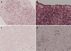
Figure 1 Histological staining of pancreatic tissue slices.
Slices derived from normal pancreas and pancreatic cancer were cultured for 3 d. A, B: Normal pancreas of good viability; C: Normal pancreas of poor viability showing massive tissue slice necrosis; D: Poorly differentiated adenocarcinoma of good viability. Hematoxylin/eosin staining. Original magnification of all tissues (100 ×).
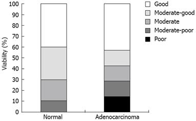
Figure 2 Viability of pancreatic (cancer) slices cultured for 3 d.
All slices were cultured for 3 d and then stained with hematoxylin/eosin. The morphology of normal pancreas (n = 10) and pancreatic adenocarcinoma (n = 6) was studied microscopically and scored by an experienced pathologist.
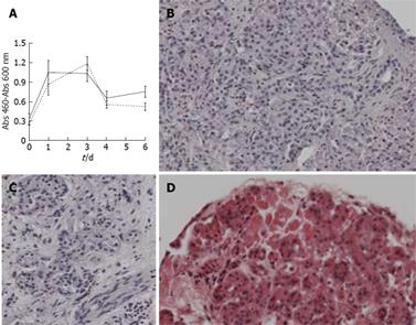
Figure 3 Viability of human pancreas tissue slices cultured ex-vivo for up to 6 d.
A: Viability of explants cultured in 1 mL DMEM (black line) or in DMEM supplemented with ITS growth factors (dashed line) was determined at day 1, 3, 4 and 6 of culturing a WST assay. All data given as the means of at least five slices ± SD; B: Normal pancreas with good viability at day 6; C: Normal pancreas of good viability at day 6 with Langerhans and nerve cells; D: Normal pancreas at day 6 with necrotic areas. Original magnification (100 ×).
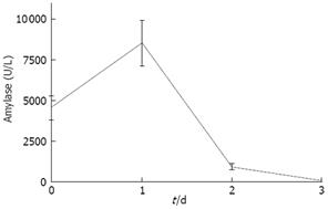
Figure 4 Amylase secretion by human pancreas tissue slices.
Human pancreatic cancer tissues were cultured ex-vivo for up to 3 d. Amount of amylase present in medium was determined using a colorimetric enzyme based assay. Data are the means ± SD (n = 12).
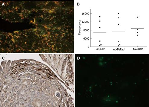
Figure 5 Transduction of pancreatic tissue slices with different viral vectors.
A: Normal human pancreatic tissue slices are incubated with 1.0 × 10⁸ Ad5.GFP and 1.0 × 10⁸ Ad5.dsRed. After 48 h presence of GFP and ds.Red was detected using a fluorescent microscope; B: Normal pancreas slices were infected with 1.0 × 10⁸ genomic copies per slice of Ad5.GFP and Ad5.dsRED, or 1.2 × 10¹⁰ viral genomes AAV2-GFP. At 48 h after infection slices were lysed, GFP and ds.Red were quantified using the Novostar; C: Pancreatic adenocarcinoma explants were transduced with Ad-GFP. At 72 h after virus addition expression of GFP was detected using an anti-GFP antibody and counterstained with hematoxylin; D: GFP expression in normal pancreas at 72 h after incubation with lentivirus-GFP.
-
Citation: Geer MAV, Kuhlmann KF, Bakker CT, Kate FJT, Elferink RPO, Bosma PJ.
Ex-vivo evaluation of gene therapy vectors in human pancreatic (cancer) tissue slices. World J Gastroenterol 2009; 15(11): 1359-1366 - URL: https://www.wjgnet.com/1007-9327/full/v15/i11/1359.htm
- DOI: https://dx.doi.org/10.3748/wjg.15.1359


