Copyright
©2009 The WJG Press and Baishideng.
World J Gastroenterol. Mar 21, 2009; 15(11): 1346-1352
Published online Mar 21, 2009. doi: 10.3748/wjg.15.1346
Published online Mar 21, 2009. doi: 10.3748/wjg.15.1346
Figure 1 Induction of apoptosis in HT-29 cells by curcumin.
Fluorescence images of HT-29 cells using Hoechst 33258 staining showed curcumin induced typical apoptotic morphological changes. A: Control; B: HT-29 cells incubated with 20 &mgr;mol/L curcumin; C: HT-29 cells incubated with 40 &mgr;mol/L curcumin; D: HT-29 cells incubated with 60 &mgr;mol/L curcumin.
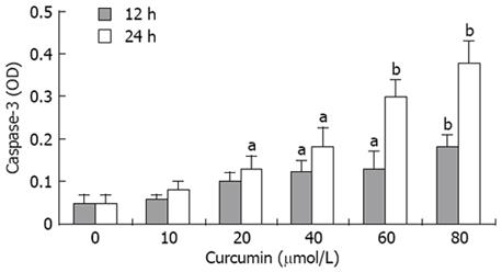
Figure 2 Caspase-3 activity was markedly increased by curcumin in a dose-dependent manner.
Each bar denotes mean ± SD (n = 3). (aP < 0.05 vs control; bP < 0.01 vs control).
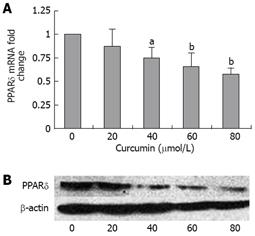
Figure 3 Curcumin inhibits the expression of PPARδ in HT-29 cells.
A: Real-time quantitative RT-PCR indicated that curcumin markedly reduced the level of PPARδ mRNA. Values are % reduction in PPARδ mRNA fold changes caused by curcumin compared with cells without curcumin treatment. Values are mean ± SD from three samples per group. GAPDH was used as an internal control; B: The effects of curcumin on the level of PPARδ protein were determined by Western blotting. β-actin was used as an internal marker. (aP < 0.05 vs control; bP < 0.01 vs control).
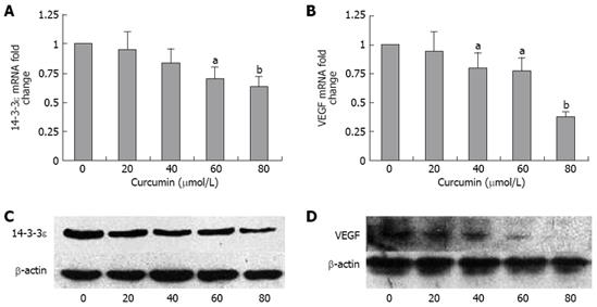
Figure 4 The expression of 14-3-3epsilon and VEGF is down-regulated by curcumin.
Real-time quantitative RT-PCR showed that curcumin decreased the mRNA levels of 14-3-3epsilon (A) and VEGF (B). Values are means ± SD from three samples per group. GAPDH was used as an internal control for quantitative RT-PCR. Western blotting was performed to determine the effects of curcumin on the expression of 14-3-3epsilon (C) and VEGF (D). β-actin was used as internal marker for Western blotting. (aP < 0.05 vs control; bP < 0.01 vs control).
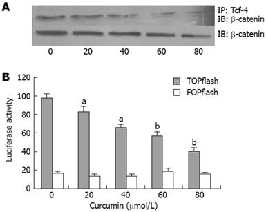
Figure 5 Curcumin inhibited β-catenin association with Tcf-4 and down-regulated the β-catenin/Tcf reporter activities in HT-29 cells.
A: Immunoprecipitation and immunoblotting results showed that curcumin inhibited β-catenin associated with Tcf-4 but had no effects on the level of β-catenin in nucleus; B: Curcumin significantly reduced TOPflash luciferase activity but had no effect on the activity of FOPflash. Values represent means ± SE of three independent experiments. (aP < 0.05 vs control; bP < 0.01 vs control).
- Citation: Wang JB, Qi LL, Zheng SD, Wang HZ, Wu TX. Curcumin suppresses PPARδ expression and related genes in HT-29 cells. World J Gastroenterol 2009; 15(11): 1346-1352
- URL: https://www.wjgnet.com/1007-9327/full/v15/i11/1346.htm
- DOI: https://dx.doi.org/10.3748/wjg.15.1346


