Copyright
©2009 The WJG Press and Baishideng.
World J Gastroenterol. Mar 21, 2009; 15(11): 1301-1314
Published online Mar 21, 2009. doi: 10.3748/wjg.15.1301
Published online Mar 21, 2009. doi: 10.3748/wjg.15.1301
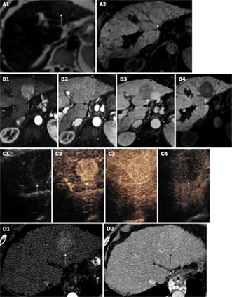
Figure 1 There is a large HCC in the left lobe of the liver with a pseudocapsule, hyper-enhancing on arterial phase and showing washout on late phases on MR and CEUS and is iso-intense in portal phases on CT and CEUS.
The pseudo-capsule enhances in the portal phase on all modalities. A1: T2 weighted scan showing slightly higher intensity HCC (arrow); A2: T1 weightted scan shows same HCC which is iso-intense (arrow); B1: MultiHance enhanced T1 weighted scan in the arterial phase showing enhancement of the HCC (arrow); B2: MultiHance enhanced T1 weighted scan in the portal phase showing iso-enhancement of the HCC (arrow); B3: MultiHance enhanced T1 weighted scan at 2 min showing contrast wash-out in the HCC (arrow); B4: MultiHance enhanced T1 weighted scan at 40 min showing hypoiintense HCC (arrow); C1: Baseline ultrasound shows iso-echoic HCC (arrow); C2: SonoVue enhanced ultrasound shows hyper-enhancing HCC (arrow) in the arterial phase; C3: SonoVue enhanced ultrasound shows iso-enhancing HCC (arrow) in the portal phase with enhancement of the pseudocapsule; C4: SonoVue enhanced ultrasound shows wash-out of the HCC (arrow) in the late phase; D1: Contrast-enhanced CT scan shows enhancement of the HCC (arrow) in the arterial phase; D2: Contrast-enhanced CT scan shows iso-enhancement of the HCC (arrow) in the portal phase with enhancement of the pseudocapsule.
- Citation: Gomaa AI, Khan SA, Leen EL, Waked I, Taylor-Robinson SD. Diagnosis of hepatocellular carcinoma. World J Gastroenterol 2009; 15(11): 1301-1314
- URL: https://www.wjgnet.com/1007-9327/full/v15/i11/1301.htm
- DOI: https://dx.doi.org/10.3748/wjg.15.1301


