Copyright
©2009 The WJG Press and Baishideng.
World J Gastroenterol. Jan 7, 2009; 15(1): 95-101
Published online Jan 7, 2009. doi: 10.3748/wjg.15.95
Published online Jan 7, 2009. doi: 10.3748/wjg.15.95
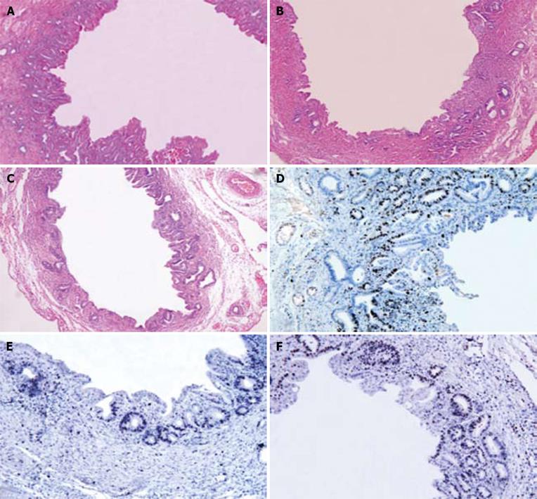
Figure 1 HE staining and immunohistochemistry for c-myc in CPC group (A, D), c-myc shRNA-3 treatment group (B, E), and c-myc shRNA-2 treatment group (C, F).
Briefly, treatment with c-myc shRNA, especially with c-myc shRNA-3, can efficaciously inhibit hyperplasia of biliary epithelium, submucosal gland, collagen fiber, and down-regulate c-myc expression (A-C, × 50; D-F, × 100).
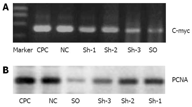
Figure 2 Real-time PCR (A) and Western blot (B) analysis of c-myc and PCNA expression in biliary duct wall.
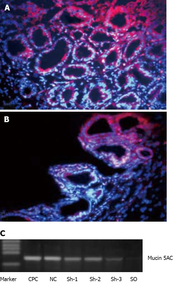
Figure 3 Immunofluorescence (A, B) and RT-PCR (C) analysis of mucin 5AC in bile duct wall.
CPC group (A), c-myc shRNA-3 treatment group (B). Briefly, treatment with c-myc shRNA-3 can result in a more prominent down-regulation of mucin 5AC expression (A and B, × 400).
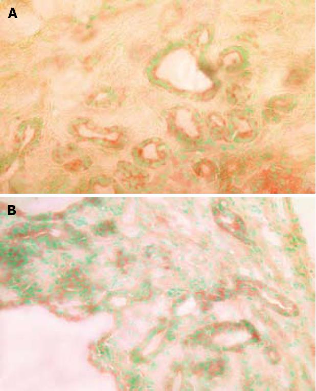
Figure 4 Enzymatic histochemistry staining of endogenous β-G (cryostat section,× 400) in CPC group (A) and c-myc shRNA-3 treatment group (B).
A notable reduction of endogenous β-G was observed in the bile duct wall following c-myc shRNA treatment.
Figure 5 Western blot analysis of procollagen III expression in biliary duct wall.
- Citation: Li FY, Cheng NS, Cheng JQ, Mao H, Jiang LS, Li N, He S. Treatment of chronic proliferative cholangitis with c-myc shRNA . World J Gastroenterol 2009; 15(1): 95-101
- URL: https://www.wjgnet.com/1007-9327/full/v15/i1/95.htm
- DOI: https://dx.doi.org/10.3748/wjg.15.95


