Copyright
©2009 The WJG Press and Baishideng.
World J Gastroenterol. Jan 7, 2009; 15(1): 38-47
Published online Jan 7, 2009. doi: 10.3748/wjg.15.38
Published online Jan 7, 2009. doi: 10.3748/wjg.15.38
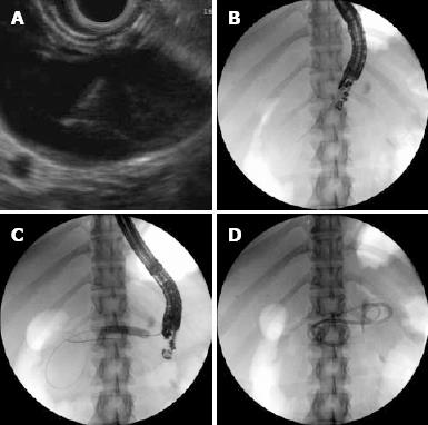
Figure 1 EUS and fluoroscopic image.
A: EUS image of pseudocyst with FNA needle; B: Fluoroscopy image of pseudocyst with FNA needle; C: Fluoroscopy image of balloon dilating the cyst gastrostomy tract; D: Fluoroscopic image of two double pigtail stents draining the pseudocyst cavity.
- Citation: Habashi S, Draganov PV. Pancreatic pseudocyst. World J Gastroenterol 2009; 15(1): 38-47
- URL: https://www.wjgnet.com/1007-9327/full/v15/i1/38.htm
- DOI: https://dx.doi.org/10.3748/wjg.15.38


