Copyright
©2008 The WJG Press and Baishideng.
World J Gastroenterol. Mar 7, 2008; 14(9): 1456-1458
Published online Mar 7, 2008. doi: 10.3748/wjg.14.1456
Published online Mar 7, 2008. doi: 10.3748/wjg.14.1456
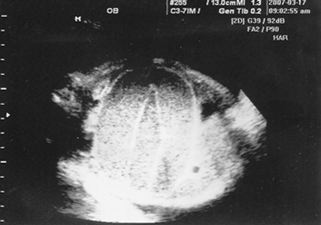
Figure 1 A fetal sonogram showing dilated bowel loops with the appearance of a ‘coffee bean sign’.
No ascites was seen in the fetal abdomen.
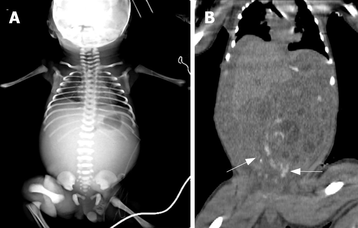
Figure 2 A: Pre-operative infantogram showing a gas shadow only in the stomach, with an absence of any distal gas shadow; B: Unenhanced abdominal CT showed meconium (arrow) in the distal small bowel, with mild fluid distension of the proximal small bowel.
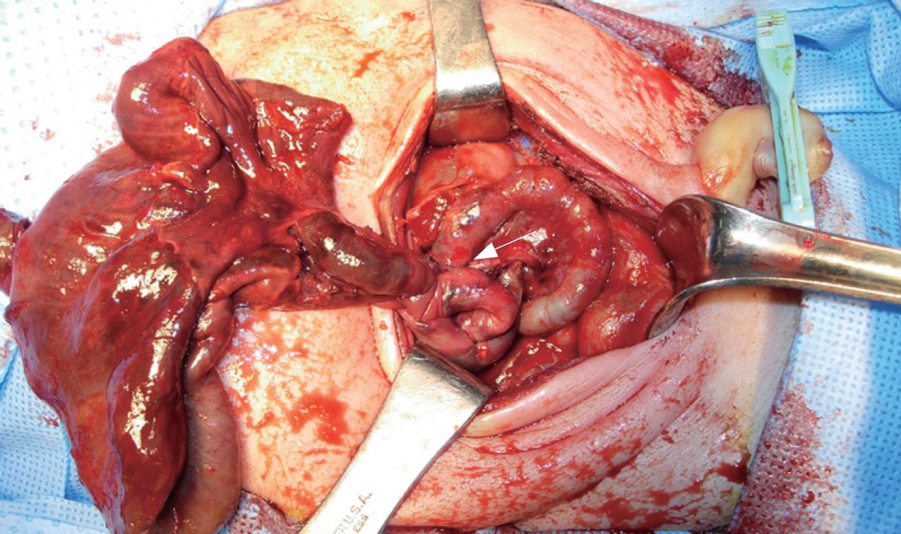
Figure 3 On laparotomy, the infant was found to have a midgut volvulus with necrosis and perforation of the small bowel.
The small bowel was found to be twisted at the level of the narrow meconium-filled distal ileum (arrow).
- Citation: Park JS, Cha SJ, Kim BG, Kim YS, Choi YS, Chang IT, Kim GJ, Lee WS, Kim GH. Intrauterine midgut volvulus without malrotation: Diagnosis from the ‘coffee bean sign’. World J Gastroenterol 2008; 14(9): 1456-1458
- URL: https://www.wjgnet.com/1007-9327/full/v14/i9/1456.htm
- DOI: https://dx.doi.org/10.3748/wjg.14.1456


