Copyright
©2008 The WJG Press and Baishideng.
World J Gastroenterol. Feb 28, 2008; 14(8): 1296-1298
Published online Feb 28, 2008. doi: 10.3748/wjg.14.1296
Published online Feb 28, 2008. doi: 10.3748/wjg.14.1296
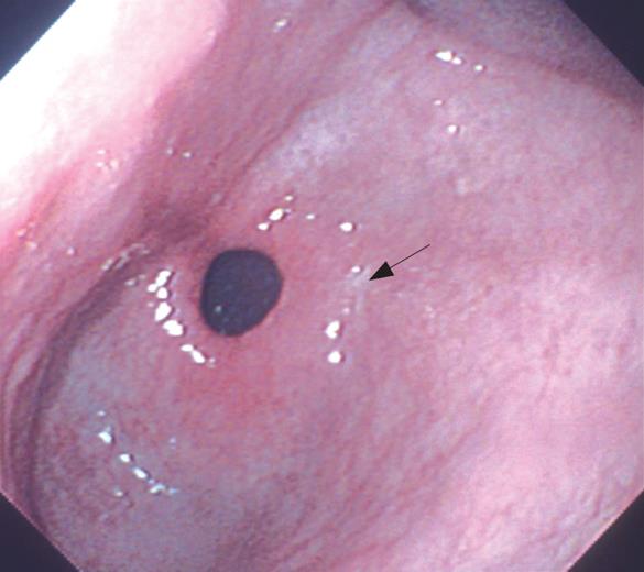
Figure 1 EGD showing a linear depressed whitish mucosal lesion of about 0.
8 cm in length (arrow).
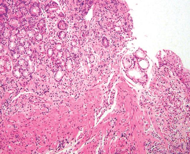
Figure 2 Photomicrograph showing squamous metaplasia (HE; original magnification, × 100).
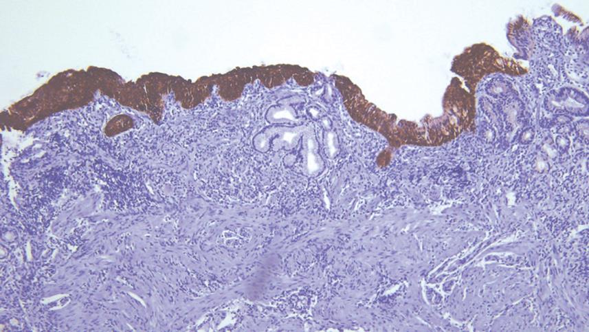
Figure 3 Photomicrograph showing an immunohistochemical demonstration of high molecular weight cytokeratin (original magnification, × 40).
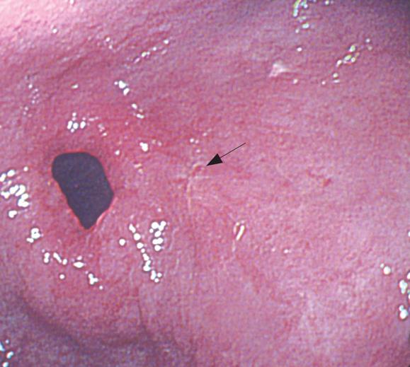
Figure 4 Follow-up EGD showing a linear depressed whitish mucosal lesion without interval change (arrow).
- Citation: Cho YS, Kim JS, Kim HK, Ji JS, Kim BW, Chae HS, Han SW, Choi KY, Chung IS. A squamous metaplasia in a gastric ulcer scar of the antrum. World J Gastroenterol 2008; 14(8): 1296-1298
- URL: https://www.wjgnet.com/1007-9327/full/v14/i8/1296.htm
- DOI: https://dx.doi.org/10.3748/wjg.14.1296


