Copyright
©2008 The WJG Press and Baishideng.
World J Gastroenterol. Feb 28, 2008; 14(8): 1182-1191
Published online Feb 28, 2008. doi: 10.3748/wjg.14.1182
Published online Feb 28, 2008. doi: 10.3748/wjg.14.1182
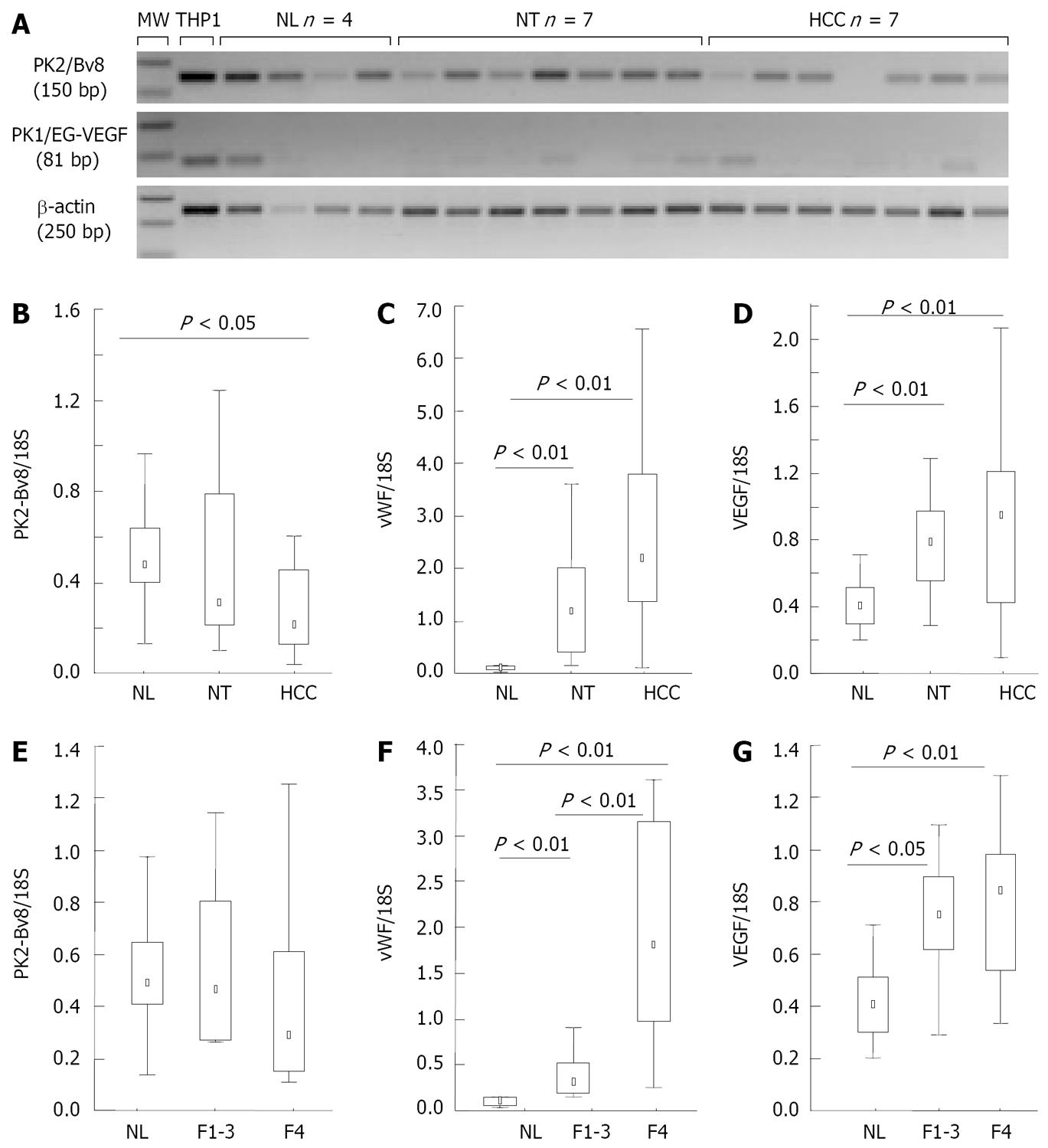
Figure 1 (A) RT-PCR analysis of PK1/EG-VEGF and PK2/Bv8, and (B-G), real-time PCR analysis of PK2/Bv8, vWF and VEGF in normal liver (NL, n = 10), non-tumorous liver (NT, n = 28), and in Hepatocellular carcinoma (HCC, n = 28).
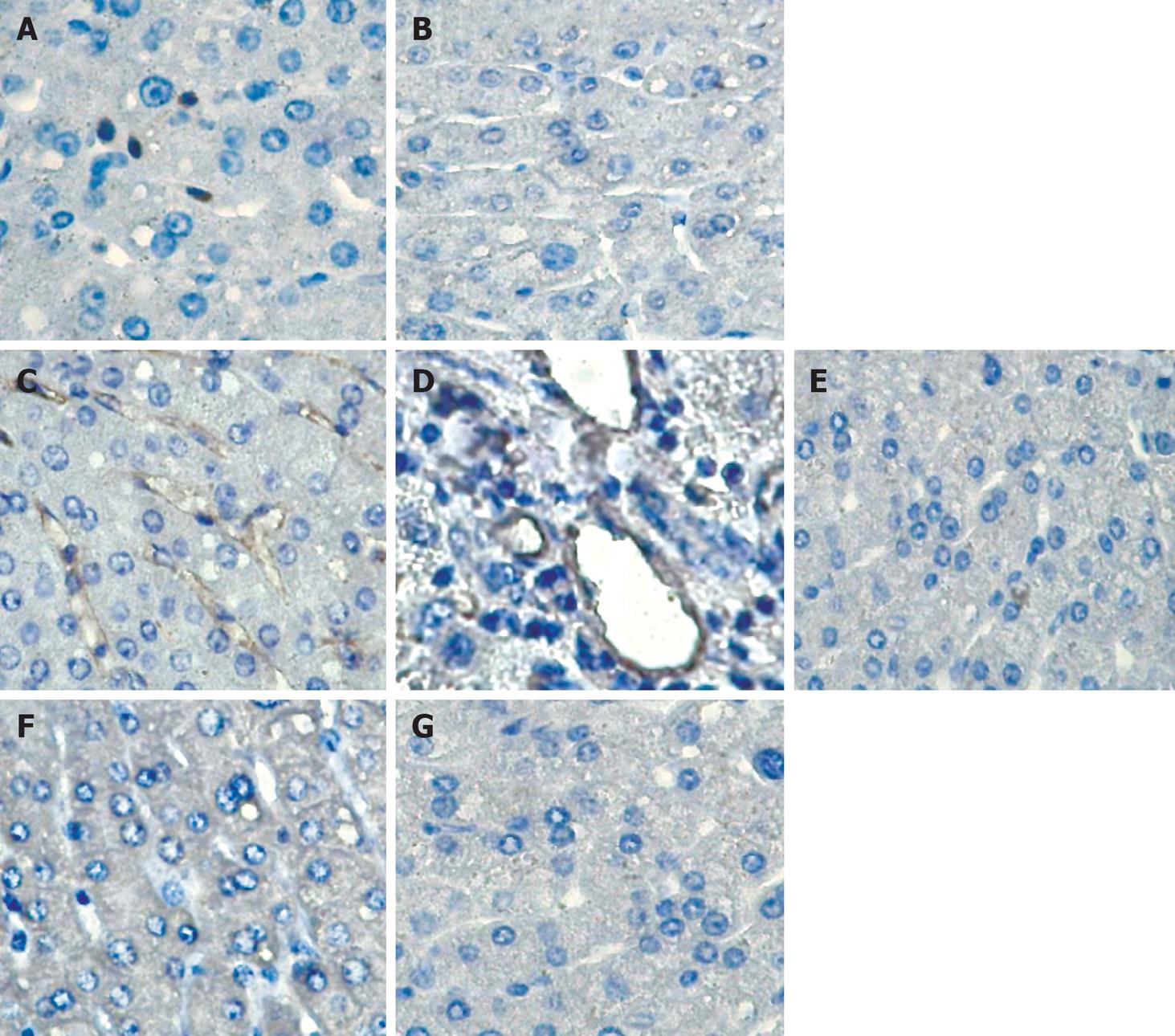
Figure 2 Immunolocalization of PK2/Bv8, CD31, CD34 and VEGF on normal liver sections.
A: Bv8; B: Bv8 + blocking peptide; C: CD31; D: CD34; E: Mouse IgG1; F: VEGF; G: Rabbit IgG.
Figure 3 (A) Expression of CD68 normal liver (NL, n = 10), non-tumorous liver (NT, n = 28), and in Hepatocellular carcinoma (HCC, n = 28) by real time PCR, and (B) immunolocalization of CD68, PK2/Bv8, and PK2/Bv8 with blocking peptide on normal liver paraffin embedded serial sections.
Figure 4 (A) Scatter plot and (B) histogram plot of CD14+enriched Kupffer cells, and (C) expression of PK1/EG-VEGF and PK2/Bv8 mRNA in isolated human liver cells by real time PCR.
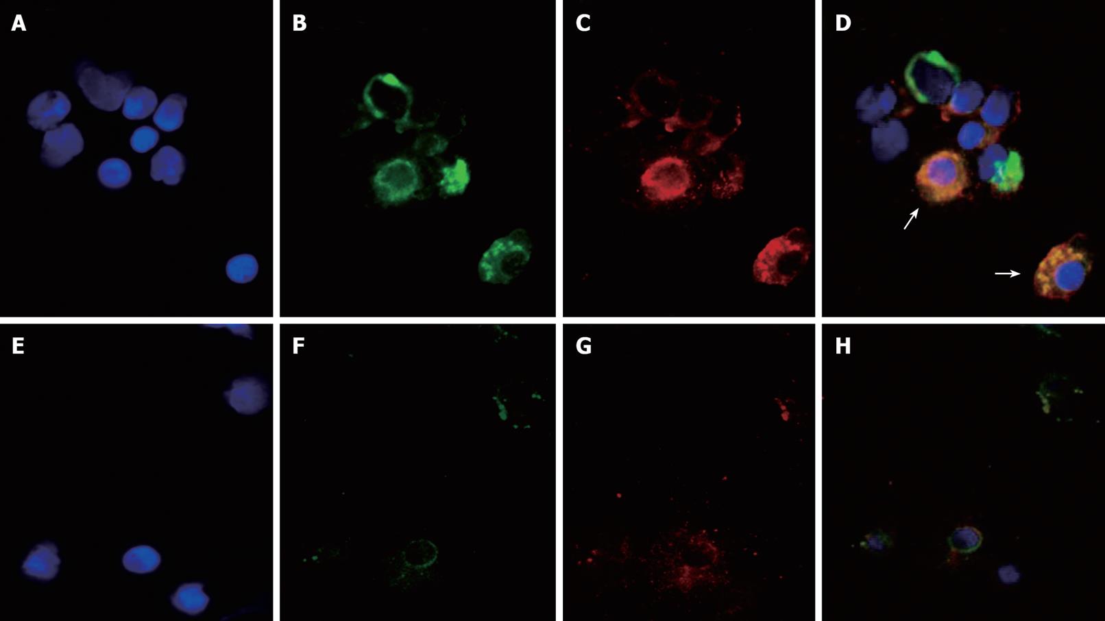
Figure 5 Immunocytochemical staining of PK2/Bv8 and CD68 on isolated liver cells enriched in CD14+ Kupffer cells.
A: DAPI; B: CD68-FITC; C: Bv8/PK2-TRITC; D: Merge; E: DAPI; F: Mouse IgG1; G: Mouse serum; H: Merge.
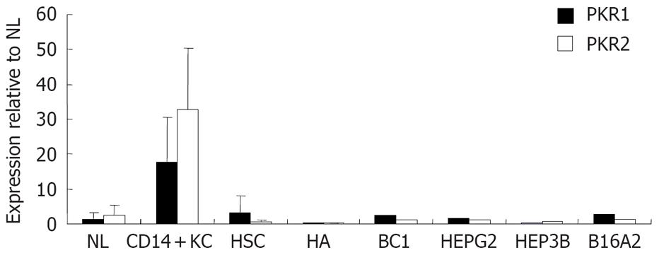
Figure 6 Expression of PKR1 and PKR2 mRNA in isolated human liver cells, by real time PCR.
- Citation: Monnier J, Piquet-Pellorce C, Feige JJ, Musso O, Clément B, Turlin B, Théret N, Samson M. Prokineticin 2/Bv8 is expressed in Kupffer cells in liver and is down regulated in human hepatocellular carcinoma. World J Gastroenterol 2008; 14(8): 1182-1191
- URL: https://www.wjgnet.com/1007-9327/full/v14/i8/1182.htm
- DOI: https://dx.doi.org/10.3748/wjg.14.1182


