Copyright
©2008 The WJG Press and Baishideng.
World J Gastroenterol. Feb 14, 2008; 14(6): 853-863
Published online Feb 14, 2008. doi: 10.3748/wjg.14.853
Published online Feb 14, 2008. doi: 10.3748/wjg.14.853
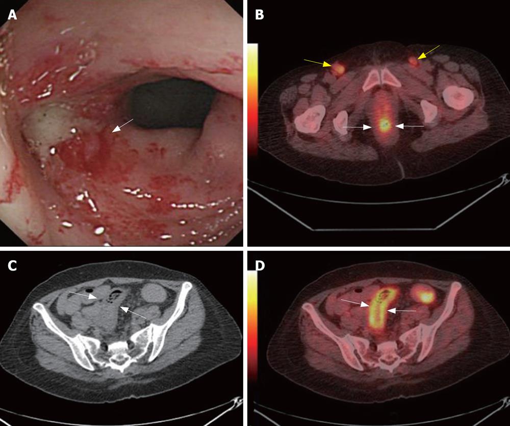
Figure 1 A 56-year-old woman was suffered from uterine cervix cancer and accepted local radiation and chemotherapy 2-mo ago.
CC found enterocolitis of sigmoid colon (white arrow, A). PET/CT detected high metabolism metastasis lymphoid nodes at both sides of the inguina (yellow arrows, B) and rectum wall was high metabolism (white arrows, B). CT showed thickening of sigmoid colon wall (white arrows, C). PET/CT illustrated sigmoid colon wall was high metabolism (white arrows, D). Biopsy followed by PET/CT revealed metastasis lymphoid nodes at both sides of the inguina. The case illustrated the potential value of PET/CTC in radiation enterocolitis.
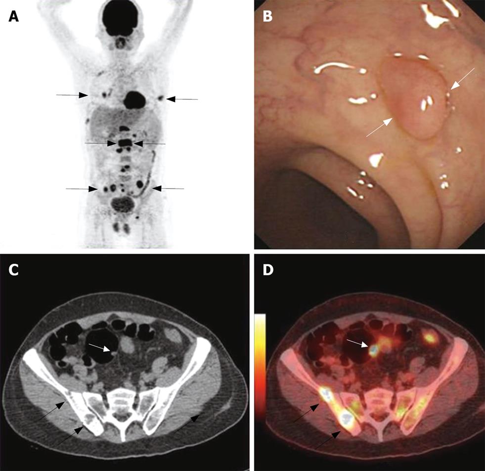
Figure 2 A 63-year-old man was detected multi metastases in whole body PET/CT scan with unknown original tumor (black arrows, A).
CC showed a polyp at the sigmoid colon (white arrows, panel B). CT detected bone destruction at the right ilium (black arrow, C) and a polyp at the sigmoid colon (white arrow, C). PET/CTC localizes the high metabolism bone destruction lesions (black arrows, D) and high metabolism polyps at sigmoid colon (white arrow, D). Histopathology follow by CC revealed a inflame polyp. The case illustrated the potential value of PET/CTC in screening colorectal polyps.
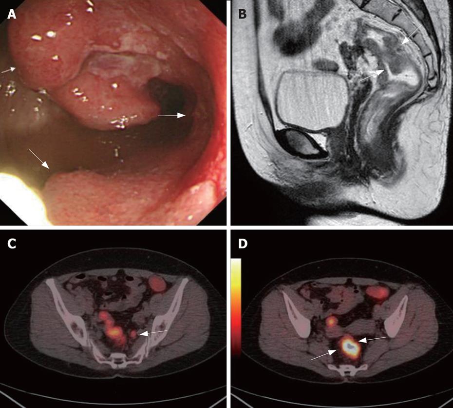
Figure 3 A 56-year-old women complained about blood stool for about 1 year.
CC showed one stenotic tumoral site in the rectum (white arrows, A). Sagittal MRC showed thickened rectum with suspected infiltration of the surrounding tissue (white arrows, B). Axial PET/CTC showed a high metabolism metastasis lymphoid node (white arrow, C). Axial PET/CTC revealed tumor sites with elevated glucose metabolism and clear circumscription (two white arrows, D). PET/CT also indicated no tumorous infiltration of the adjacent tissue and no distant metastases. This was later verified by histopathology. The case illustrated the value of VC in CRC TNM staging.
- Citation: Sun L, Wu H, Guan YS. Colonography by CT, MRI and PET/CT combined with conventional colonoscopy in colorectal cancer screening and staging. World J Gastroenterol 2008; 14(6): 853-863
- URL: https://www.wjgnet.com/1007-9327/full/v14/i6/853.htm
- DOI: https://dx.doi.org/10.3748/wjg.14.853


