Copyright
©2008 The WJG Press and Baishideng.
World J Gastroenterol. Feb 7, 2008; 14(5): 758-763
Published online Feb 7, 2008. doi: 10.3748/wjg.14.758
Published online Feb 7, 2008. doi: 10.3748/wjg.14.758
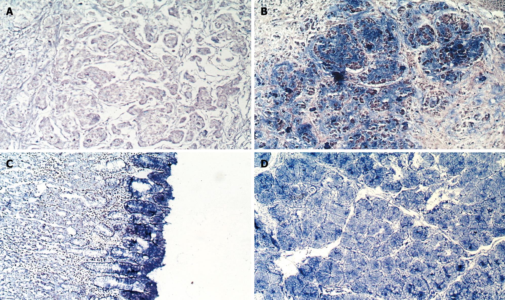
Figure 1 In situ hybridization analysis of GCRG123 in gastric mucosa.
GCRG123 showed low expression (blue precipitates restricted to the cytoplasm) in intestinal-type adenocarcinoma (A) and normal gastric glands (C, left region); high expression in signet-ring cell carcinoma (B); normal epithelia (C); and normal pyloric glands (D). NBT/BCIP was used as an alkaline phosphatase substrate (magnification was 10 × 10).
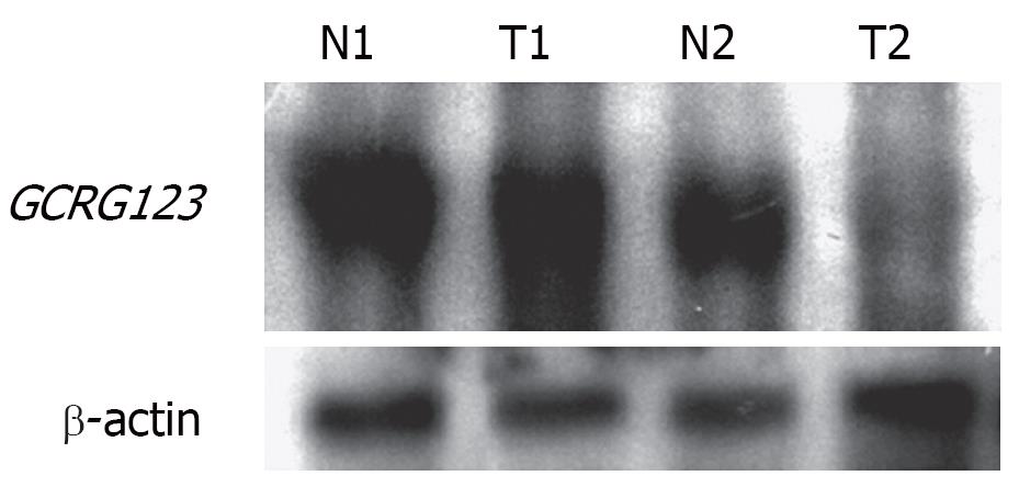
Figure 2 Northern blotting of GCRG123 in gastric tissues.
The expression of β-actin served as an internal control. Total RNA, 1 &mgr;g per lane, was separated on a 1% agarose/formaldehyde gel. After blotting onto a nylon membrane, the membrane was probed with digoxigenin-labeled anti-sense GCRG123 cRNA. N, normal gastric tissue; T, tumor; 1, signet-ring cell carcinoma; 2, intestinal-type adenocarcinoma.
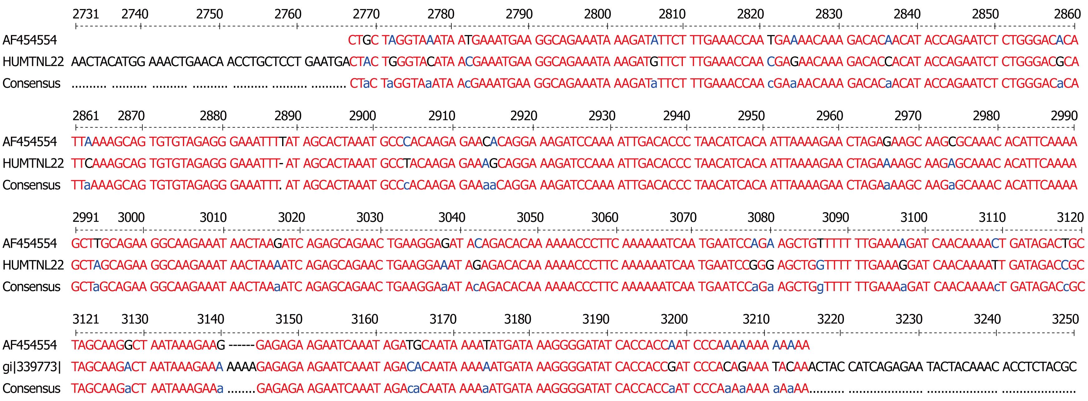
Figure 3 Multalin analysis of GCRG123 (AF454554) and L1 (HUMTNL22).
Red font represents high consensus, blue or black font represents low consensus.
-
Citation: Wang GS, Wang MW, Wu BY, Yang XY, Wang WH, You WD.
LINE-1 family memberGCRG123 gene is up-regulated in human gastric signet-ring cell carcinoma. World J Gastroenterol 2008; 14(5): 758-763 - URL: https://www.wjgnet.com/1007-9327/full/v14/i5/758.htm
- DOI: https://dx.doi.org/10.3748/wjg.14.758


