Copyright
©2008 The WJG Press and Baishideng.
World J Gastroenterol. Dec 21, 2008; 14(47): 7252-7255
Published online Dec 21, 2008. doi: 10.3748/wjg.14.7252
Published online Dec 21, 2008. doi: 10.3748/wjg.14.7252
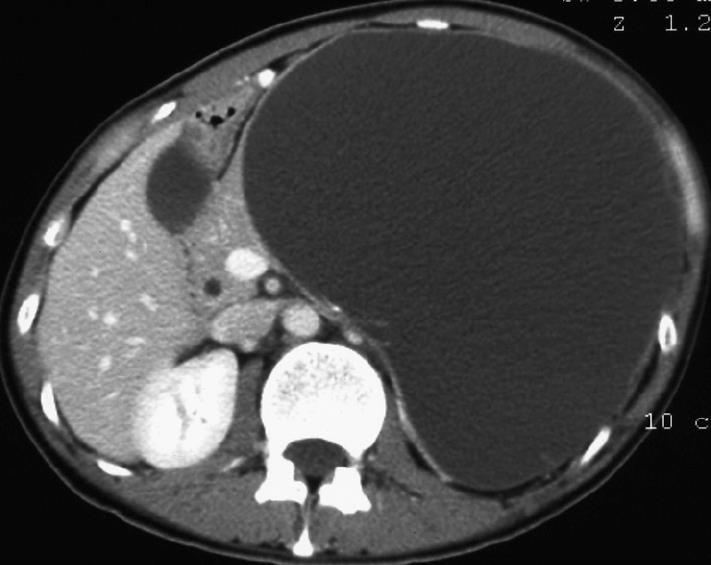
Figure 1 Abdominal computed tomography scan showing a huge cyst measuring 18 cm in diameter.
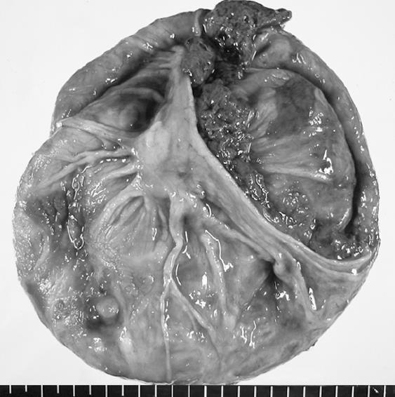
Figure 2 Macroscopic view of the cystic tumor.
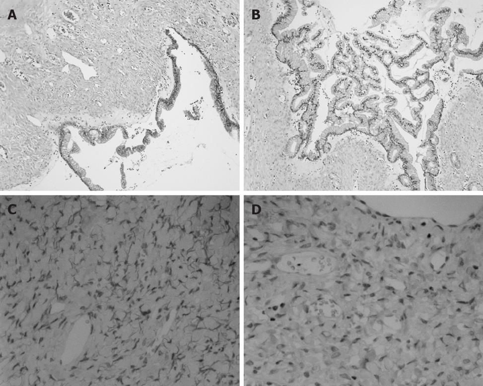
Figure 3 Microscopic findings of the cystic tumor.
A: Columnar, mucin-producing epithelium with underlying ovarian-type stroma (HE, × 100); B: The epithelium had a focal papillary architecture (HE, × 100); C: Positive staining in the stromal cell nuclei for progesterone receptor (× 400); D: The estrogen receptor of stromal cell nuclei (× 400).
- Citation: Ikuta SI, Aihara T, Yasui C, Iida H, Yanagi H, Mitsunobu M, Kakuno A, Yamanaka N. Large mucinous cystic neoplasm of the pancreas associated with pregnancy. World J Gastroenterol 2008; 14(47): 7252-7255
- URL: https://www.wjgnet.com/1007-9327/full/v14/i47/7252.htm
- DOI: https://dx.doi.org/10.3748/wjg.14.7252


