Copyright
©2008 The WJG Press and Baishideng.
World J Gastroenterol. Dec 7, 2008; 14(45): 7005-7008
Published online Dec 7, 2008. doi: 10.3748/wjg.14.7005
Published online Dec 7, 2008. doi: 10.3748/wjg.14.7005
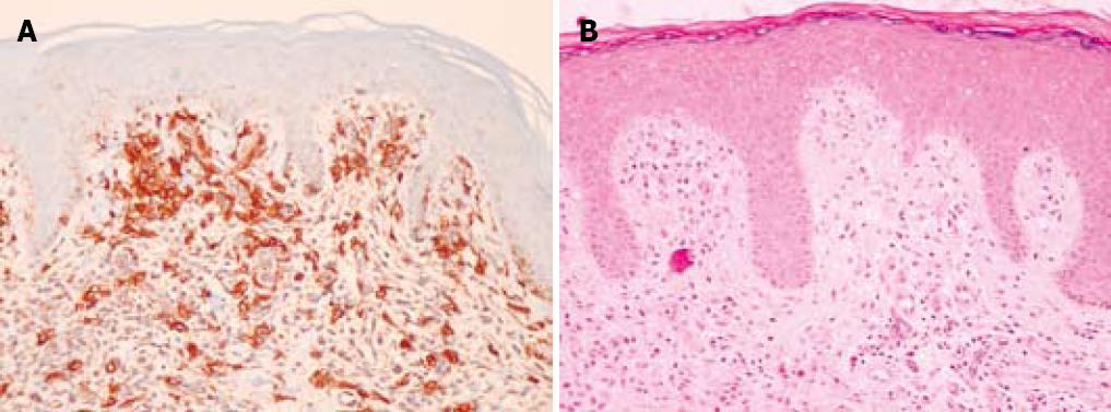
Figure 1 The patient subsequently had maculopapular rashes which on biopsy showed increased numbers of dermal mast cells highlighted by c-kit (CD 117) immunoperoxidase staining consistent with that of dermal mastocytosis.
The haematoxylin and eosin stain (HE) is also included.
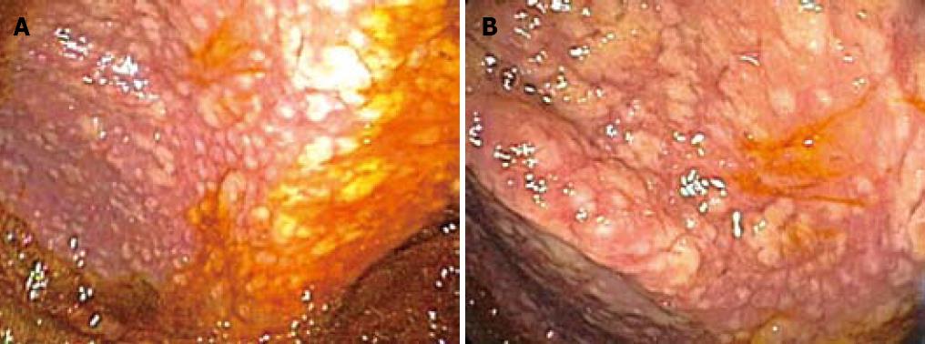
Figure 2 On colonoscopy a mucus type material adherent to the mucosa was present in the right colon along with a slightly raised appearance of the mucosa through areas of the transverse and right colon.
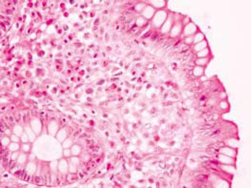
Figure 3 The biopsy in these colonic regions showed an increased number of mast cells with recruited eosinophils in the lamina propria (HE).
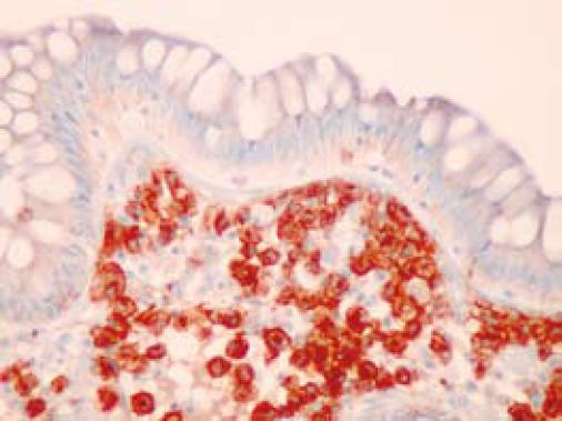
Figure 4 The mast cells are highlighted by positive c-kit staining.
- Citation: Lee JK, Whittaker SJ, Enns RA, Zetler P. Gastrointestinal manifestations of systemic mastocytosis. World J Gastroenterol 2008; 14(45): 7005-7008
- URL: https://www.wjgnet.com/1007-9327/full/v14/i45/7005.htm
- DOI: https://dx.doi.org/10.3748/wjg.14.7005


