Copyright
©2008 The WJG Press and Baishideng.
World J Gastroenterol. Nov 28, 2008; 14(44): 6808-6816
Published online Nov 28, 2008. doi: 10.3748/wjg.14.6808
Published online Nov 28, 2008. doi: 10.3748/wjg.14.6808
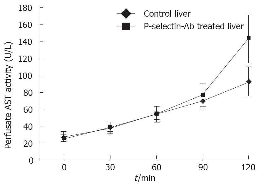
Figure 1 Perfusate aspartate aminotransferase (AST) activity in isolated-blood-perfused control and P-selectin-Ab treated rat livers after 6 h of cold ischemia.
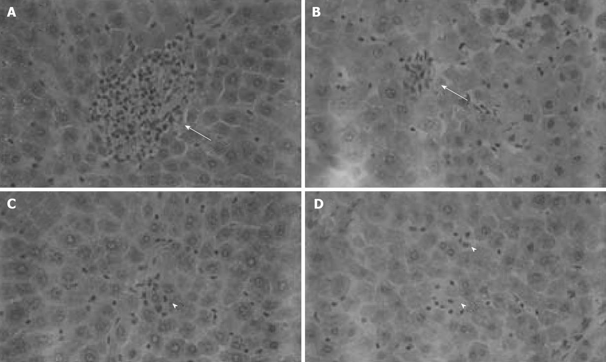
Figure 2 Histological analysis of isolated-blood-perfused control and P-selectin Ab-treated rat liver sections at 120 min perfusion after 6 h of cold ischemia (HE × 400).
A, B: Point necroses in livers of control and P-selectin Ab-treated livers at 120 min perfusion (solid arrows); C, D: Inflammation in both control and P-selectin Ab-treated livers at 120 min of perfusion (arrow heads).
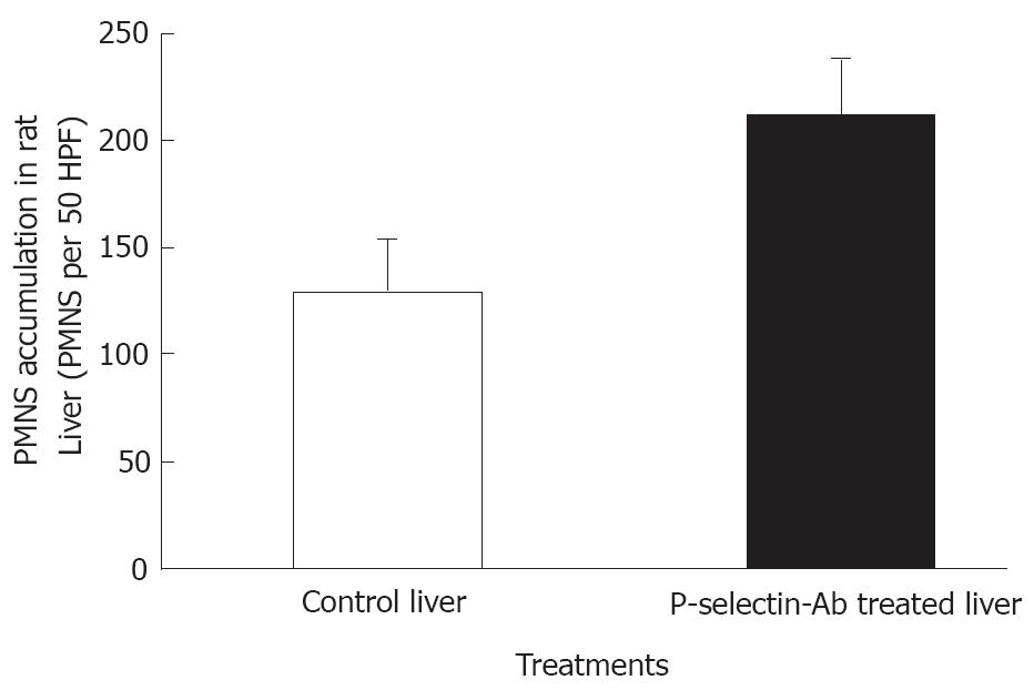
Figure 3 Accumulation of polymorphonuclear leukocytes (PMNs) in isolated-blood-perfused control and P-selectin Ab-treated rat livers at 120 min perfusion after 6 h of cold ischemia.
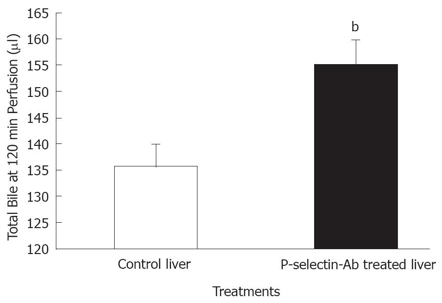
Figure 4 Bile production by isolated-blood-perfused control and P-selectin Ab-treated rat livers after 6 h of cold ischemia.
bP = 0.009 vs control liver.
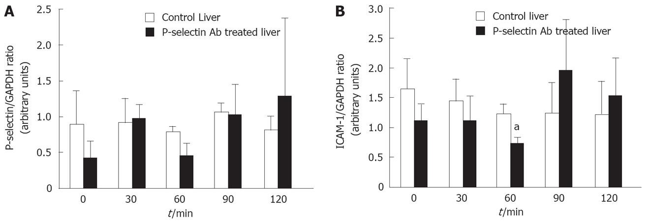
Figure 5 Semi-quantitative RT-PCR analysis of P-selectin and ICAM-1 mRNA levels in isolated-blood-perfused control and P-selectin Ab-treated rat livers after 6 h of cold ischemia.
aP < 0.05 vs control liver.
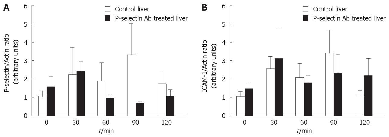
Figure 6 Western blot analysis of P-selectin and ICAM-1 protein levels in isolated-blood-perfused control and P-selectin Ab-treated rat livers after 6 h of cold ischemia.
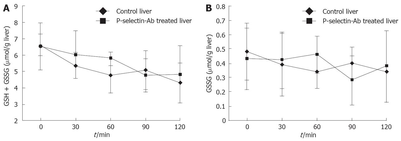
Figure 7 Reduced (GSH + GSSG) and oxidized (GSSG) glutathione levels in isolated-blood-perfused control and P-selectin Ab-treated rat livers after 6 h of cold ischemia.
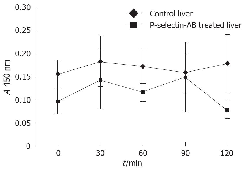
Figure 8 ELISA analysis of nuclear p65 as a measure of liver NF-κB activation in isolated-blood-perfused control and P-selectin Ab-treated rat livers after 6 h of cold ischemia.
- Citation: Wyllie S, Barshes NR, Gao FQ, Karpen SJ, Goss JA. Failure of P-selectin blockade alone to protect the liver from ischemia-reperfusion injury in the isolated blood-perfused rat liver. World J Gastroenterol 2008; 14(44): 6808-6816
- URL: https://www.wjgnet.com/1007-9327/full/v14/i44/6808.htm
- DOI: https://dx.doi.org/10.3748/wjg.14.6808


