Copyright
©2008 The WJG Press and Baishideng.
World J Gastroenterol. Nov 21, 2008; 14(43): 6757-6759
Published online Nov 21, 2008. doi: 10.3748/wjg.14.6757
Published online Nov 21, 2008. doi: 10.3748/wjg.14.6757
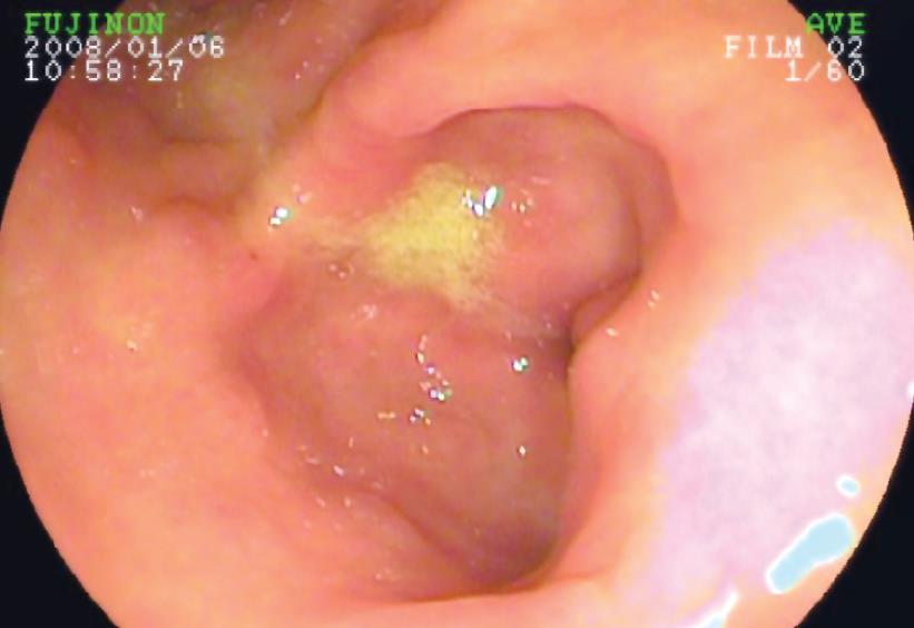
Figure 1 Submucosal lesion in the prepyloric posterior gastric wall.
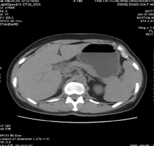
Figure 2 CT scan shows thickness of posterior gastric wall, measuring 1 cm × 1.
5 cm in the lower body of the stomach.
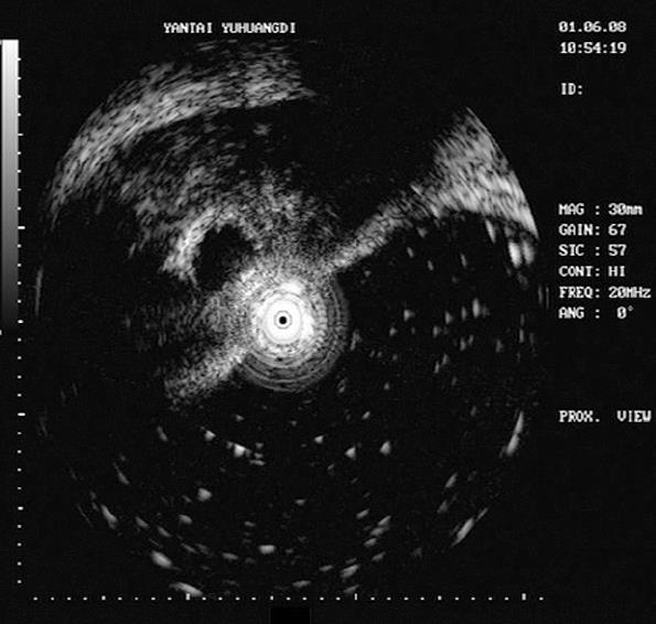
Figure 3 EUS reveals a lesion of 1.
5 cm in diameter, with low and high complex echo-genicity, located within either the third or the fourth echo-layers.
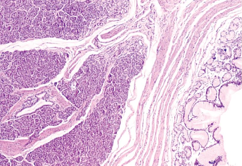
Figure 4 Heterotopic pancreatic tissue with fully developed acini and ducts.
Islet is not found.
- Citation: Jiang LX, Xu J, Wang XW, Zhou FR, Gao W, Yu GH, Lv ZC, Zheng HT. Gastric outlet obstruction caused by heterotopic pancreas: A case report and a quick review. World J Gastroenterol 2008; 14(43): 6757-6759
- URL: https://www.wjgnet.com/1007-9327/full/v14/i43/6757.htm
- DOI: https://dx.doi.org/10.3748/wjg.14.6757


