Copyright
©2008 The WJG Press and Baishideng.
World J Gastroenterol. Nov 21, 2008; 14(43): 6753-6756
Published online Nov 21, 2008. doi: 10.3748/wjg.14.6753
Published online Nov 21, 2008. doi: 10.3748/wjg.14.6753
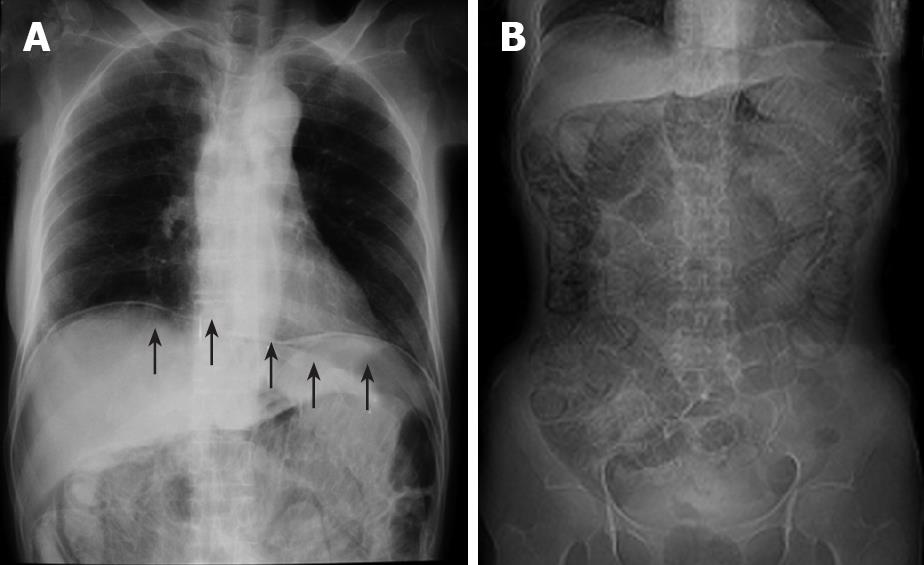
Figure 1 Abdominal roentgenogram.
A: Massive intraperitoneal free air (arrows); B: dilatation of the intestine and circular collection of intestinal gas.
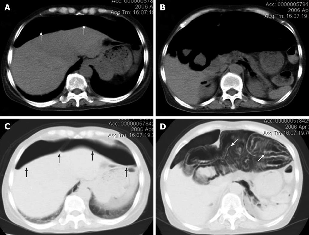
Figure 2 Abdominal CT scan images.
Massive intraperitoneal free air extending throughout the abdominal cavity (white arrows, A and B), and a stranded appearance of air collection within the bowel wall detected in the lung window setting, which extended from the jejunum to the splenic flexure of the colon. This indicated that the air was present within the bowel wall (white arrows, C and D). Black arrows indicate massive intraperitoneal air (C).
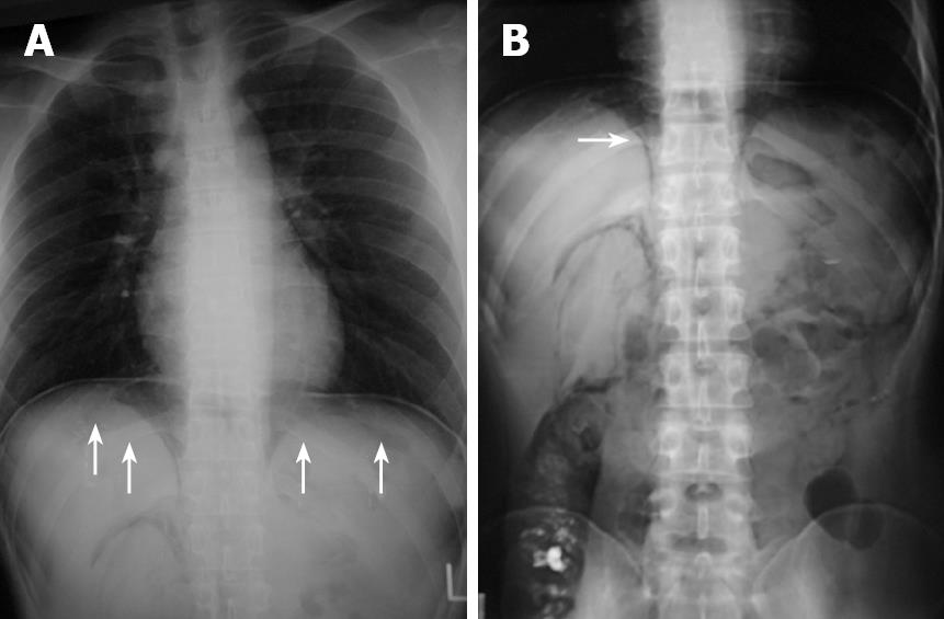
Figure 3 Roentgenogram of chest and abdomen.
It revealed massive intra-abdominal free air (arrows, A) and pneunoretroperitoneum (arrow, B).
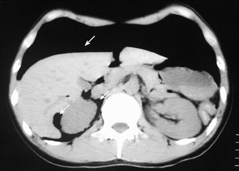
Figure 4 Abdominal CT scan, revealing massive peritoneal free air (arrows) that extended throughout the entire abdomen.
- Citation: Sakurai Y, Hikichi M, Isogaki J, Furuta S, Sunagawa R, Inaba K, Komori Y, Uyama I. Pneumatosis cystoides intestinalis associated with massive free air mimicking perforated diffuse peritonitis. World J Gastroenterol 2008; 14(43): 6753-6756
- URL: https://www.wjgnet.com/1007-9327/full/v14/i43/6753.htm
- DOI: https://dx.doi.org/10.3748/wjg.14.6753


