Copyright
©2008 The WJG Press and Baishideng.
World J Gastroenterol. Nov 21, 2008; 14(43): 6748-6749
Published online Nov 21, 2008. doi: 10.3748/wjg.14.6748
Published online Nov 21, 2008. doi: 10.3748/wjg.14.6748
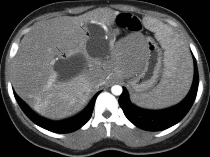
Figure 1 CT scan illustrating saccular dilations (arrows) of right and left hepatic ducts.
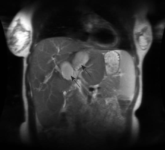
Figure 2 MRCP demonstrating saccular dilation (arrows) of the right and left hepatic ducts.
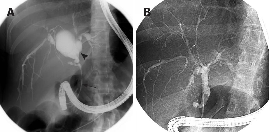
Figure 3 ERCP.
A: Initial cholangiogram showing dilated extrahepatic ducts consistent with diagnosis of type IV choledochal cysts (arrowhead), and 7 cm common duct stricture (arrow); B: Final balloon occlusion cholangiogram performed one year after stenting showing resolution of the common duct stricture and cystic biliary dilation. The intrahepatic ducts are diffusely irregular.
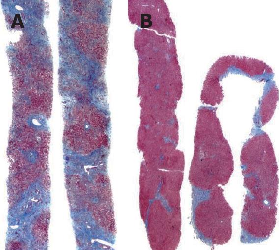
Figure 4 Liver biopsy.
A: Pretreatment Trichome stain showed cirrhosis; B: Trichrome stain one year later following biliary stenting showed marked improvement in degree of fibrosis.
- Citation: Goldwire FW, Norris WE, Koff JM, Goodman ZD, Smith MT. An unusual presentation of primary sclerosing cholangitis. World J Gastroenterol 2008; 14(43): 6748-6749
- URL: https://www.wjgnet.com/1007-9327/full/v14/i43/6748.htm
- DOI: https://dx.doi.org/10.3748/wjg.14.6748


