Copyright
©2008 The WJG Press and Baishideng.
World J Gastroenterol. Nov 21, 2008; 14(43): 6673-6680
Published online Nov 21, 2008. doi: 10.3748/wjg.14.6673
Published online Nov 21, 2008. doi: 10.3748/wjg.14.6673
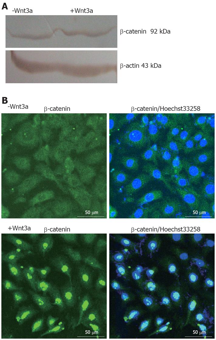
Figure 1 Effects of Wnt3a on β-catenin expression, its subcellular localization, and induction of typical Wnt target genes.
A: Stimulation of WB-F344 cells with 160 ng/mL Wnt3a for 1 d revealing a slight increase in β-catenin protein level as shown by Western blot analysis and densitometric analysis; B: Immunocytochemistry analysis of β-catenin exhibiting perinuclear staining for β-catenin in unstimulated WB-F344 cells (upper panels), whereas addition of 160 ng/mL Wnt3a for 1 d (lower panels) showing clear nuclear staining for β-catenin. Immunofluorescence was performed using a polyclonal antibody against β-catenin (left panels). In addition, nuclei of WB-F344 cells were stained with Hoechst33258 (right panels). Scale bars: 50 μm.
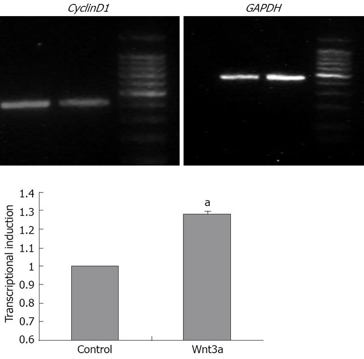
Figure 2 The mRNA expression levels of the known Wnt target genes CyclinD1 was semi-quantified at day 1 during stimulation with Wnt3a (160 ng/mL) and normalized to the expression levels in untreated WB-F344 cells (set as 100%).
The mRNA expression level of CyclinD1, one of the known Wnt target genes in WB-F344 cells after stimulation by Wnt3a (160 ng/mL) for 1 d was semi-quantified by RT-PCR and the results was normalized to the expression levels in untreated WB-F344 cells (set as 1). CyclinD1 was upregulated under stimulating conditions on day 1. For quantification, CyclinD1 mRNA was scanned by densitometric analysis and normalized to GAPDH. Data are presented as mean ± SD of triplicate experiments. aP < 0.05 in comparison with un-treated cells.
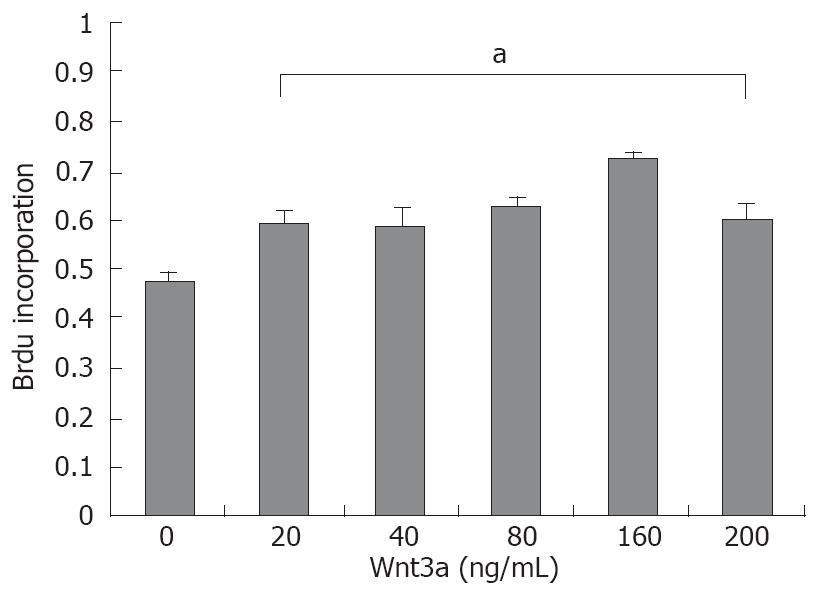
Figure 3 Proliferation of WB-F344 cells upon treatment with Wnt3a measured by Brdu incorporation assay.
The proliferation of WB-F344 cells was significantly enhanced by stimulation with recombinant Wnt3a for 1 d Data are presented as mean ± SD. aP < 0.05 vs untreated WB-F344 cells.
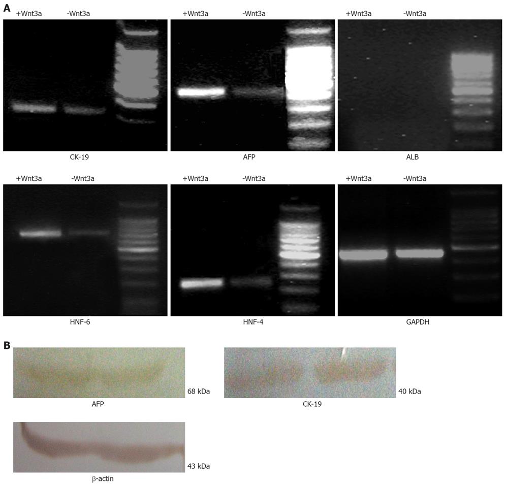
Figure 4 RT-PCR and Western blot analysis of differentiated WB-F344 cells treated or untreated with Wnt3a.
A: Wnt3a-treated WB-F344 cells expressing two phenotypic markers (CK-19 and AFP) and two hepatic nuclear factors (HNF4α and HNF-6) at mRNA level with untreated WB-F344 cells as controls; B: Wnt3a-treated cells expressing two phenotypic markers (CK-19 and AFP) at protein level.
-
Citation: Zhang Y, Li XM, Zhang FK, Wang BE. Activation of canonical Wnt signaling pathway promotes proliferation and self-renewal of rat hepatic oval cell line WB-F344
in vitro . World J Gastroenterol 2008; 14(43): 6673-6680 - URL: https://www.wjgnet.com/1007-9327/full/v14/i43/6673.htm
- DOI: https://dx.doi.org/10.3748/wjg.14.6673


