Copyright
©2008 The WJG Press and Baishideng.
World J Gastroenterol. Nov 14, 2008; 14(42): 6593-6595
Published online Nov 14, 2008. doi: 10.3748/wjg.14.6593
Published online Nov 14, 2008. doi: 10.3748/wjg.14.6593
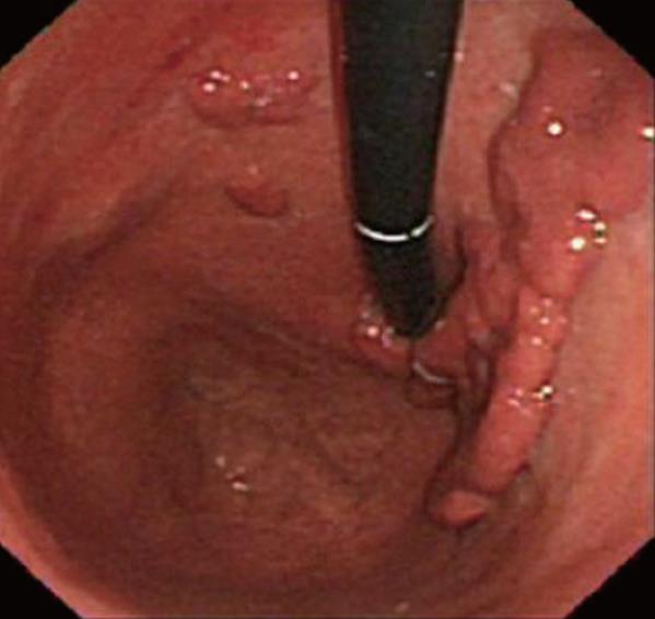
Figure 1 Endoscopic appearance of the giant FGP and the two small satellite polyps.
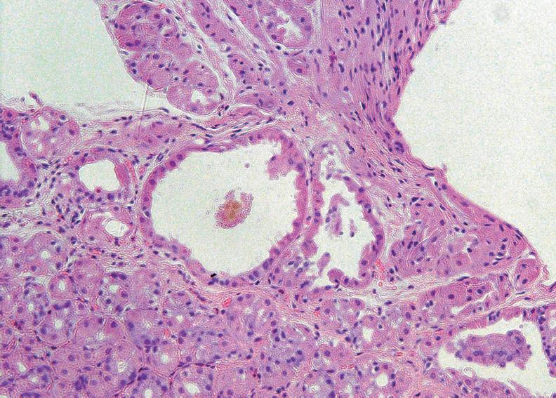
Figure 2 Fundic gland polyp biopsy showing numerous fundic glands, several of which are cystically dilated (HE, ×100).
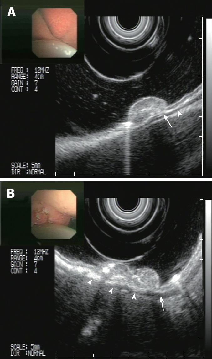
Figure 3 EUS images of the gastric polyp.
A: The polyp involves the mucosa without reaching the submucosa (arrow) or muscularis propria (arrowhead); B: Mixed echogenicity of the polyp (EUS view at a different level).
- Citation: Hajj IIE, Hawchar M, Soweid A, Maasri K, Tawil A, Barada KA. Giant sporadic fundic gland polyp: Endoscopic and endosonographic features and management. World J Gastroenterol 2008; 14(42): 6593-6595
- URL: https://www.wjgnet.com/1007-9327/full/v14/i42/6593.htm
- DOI: https://dx.doi.org/10.3748/wjg.14.6593


