Copyright
©2008 The WJG Press and Baishideng.
World J Gastroenterol. Nov 14, 2008; 14(42): 6578-6580
Published online Nov 14, 2008. doi: 10.3748/wjg.14.6578
Published online Nov 14, 2008. doi: 10.3748/wjg.14.6578
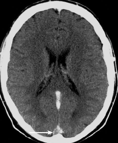
Figure 1 MRI post contrast with “Empty Delta” sign indicating a cerebral venous thrombosis.
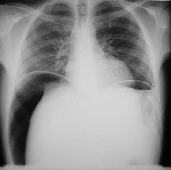
Figure 2 Erect chest X-ray demonstrating free air under the right hemidiaphragm.
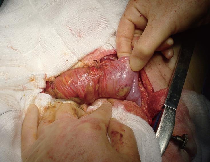
Figure 3 Punched-out ulcer at laparotomy.
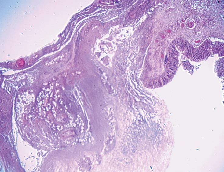
Figure 4 Microscopic specimen demonstrating loss of normal mucosa.
- Citation: Dowling CM, Hill AD, Malone C, Sheehan JJ, Tormey S, Sheahan K, McDermott E, O’Higgins NJ. Colonic perforation in Behçet’s syndrome. World J Gastroenterol 2008; 14(42): 6578-6580
- URL: https://www.wjgnet.com/1007-9327/full/v14/i42/6578.htm
- DOI: https://dx.doi.org/10.3748/wjg.14.6578


