Copyright
©2008 The WJG Press and Baishideng.
World J Gastroenterol. Nov 14, 2008; 14(42): 6518-6524
Published online Nov 14, 2008. doi: 10.3748/wjg.14.6518
Published online Nov 14, 2008. doi: 10.3748/wjg.14.6518
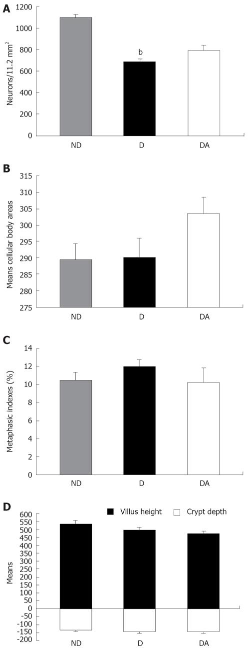
Figure 1 Quantitative and morphometric neurons meyenteric stained of myosin-V analyses.
A: Number of myenteric neurons myosin-V stained quantified in 11.2 mm2 in the jejunum of rats from groups: ND, D and DA, mean ± SE (n = 5). bP < 0.001, vs the corresponding values in group ND; B: Means (μm2) of neuronal area of myosin-V-stained myenteric neurons in animals from groups: ND, D and DA, mean ± SE (n = 5). There were no significant differences when comparing the three groups by Kruskal-Wallis test; C: IMs (%) of animals from groups: ND, D and DA, mean ± SE (n = 5). There were no significative differences when comparing the three groups by test of Tukey; D: Villus height (μm) and crypt depth (μm) in the jejunal mucosa of animals from groups: ND, D and DA, mean ± SE (n = 5). There were no significant differences when comparing the three groups by test of Tukey.
- Citation: Freitas P, Natali MRM, Pereira RVF, Neto MHM, Zanoni JN. Myenteric neurons and intestinal mucosa of diabetic rats after ascorbic acid supplementation. World J Gastroenterol 2008; 14(42): 6518-6524
- URL: https://www.wjgnet.com/1007-9327/full/v14/i42/6518.htm
- DOI: https://dx.doi.org/10.3748/wjg.14.6518


