Copyright
©2008 The WJG Press and Baishideng.
World J Gastroenterol. Jan 28, 2008; 14(4): 644-646
Published online Jan 28, 2008. doi: 10.3748/wjg.14.644
Published online Jan 28, 2008. doi: 10.3748/wjg.14.644
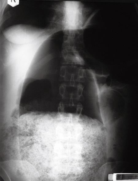
Figure 1 Plain X-ray of the abdomen showing a dilated intestinal loop occupying more than half of the abdominal cavity filled with fecal masses.
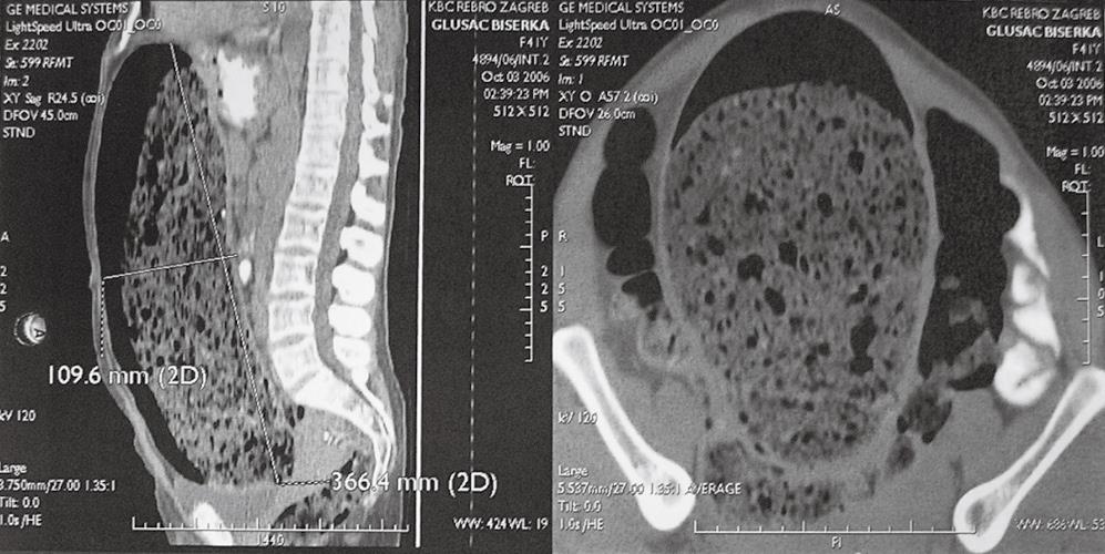
Figure 2 Abdominal CT showing a dilated intestine from the diaphargm to the pelvis, measuring 36 cm in length with a diameter of 11 cm dislocating normal intestine laterally.
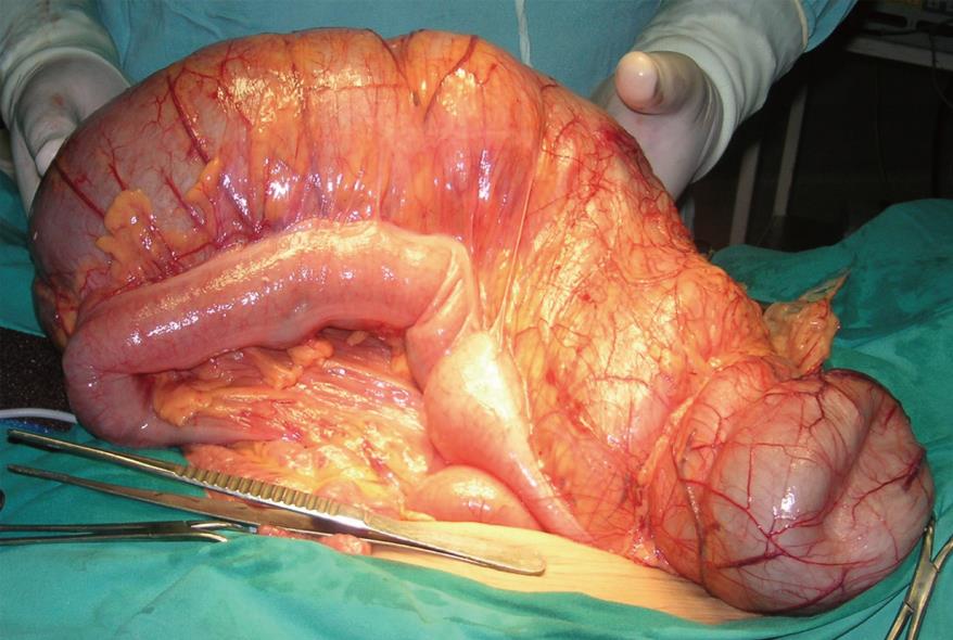
Figure 3 Operative view of normal transversal colon and the distended tubular colonic duplication.
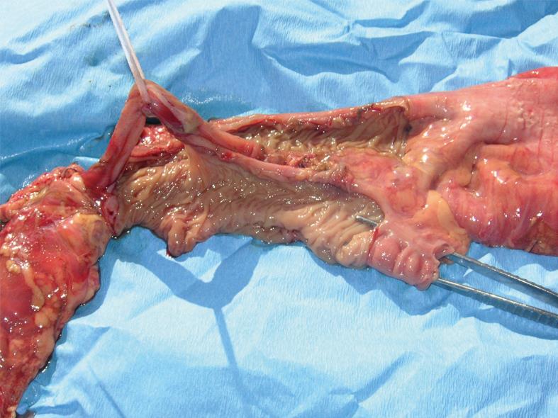
Figure 4 Surgical specimen showing part of the normal ascending and transversal colon together with a tubular duplication with all layers of the colonic wall dividing these structures.
- Citation: Kekez T, Augustin G, Hrstic I, Smud D, Majerovic M, Jelincic Z, Kinda E. Colonic duplication in an adult who presented with chronic constipation attributed to hypothyroidism. World J Gastroenterol 2008; 14(4): 644-646
- URL: https://www.wjgnet.com/1007-9327/full/v14/i4/644.htm
- DOI: https://dx.doi.org/10.3748/wjg.14.644


