Copyright
©2008 The WJG Press and Baishideng.
World J Gastroenterol. Oct 21, 2008; 14(39): 6078-6082
Published online Oct 21, 2008. doi: 10.3748/wjg.14.6078
Published online Oct 21, 2008. doi: 10.3748/wjg.14.6078
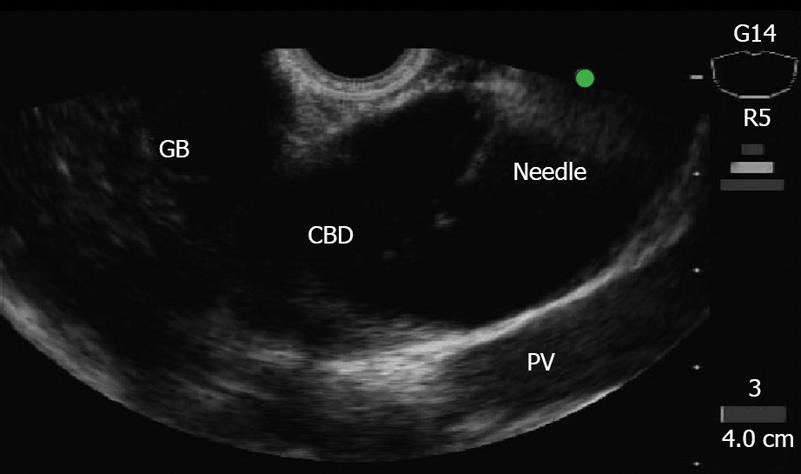
Figure 1 Convex echoendoscope clearly depicts the extrahepatic bile duct (green) (patient 3).
GB: Gallbladder; CBD: Common bile duct; PV: Portal vein.
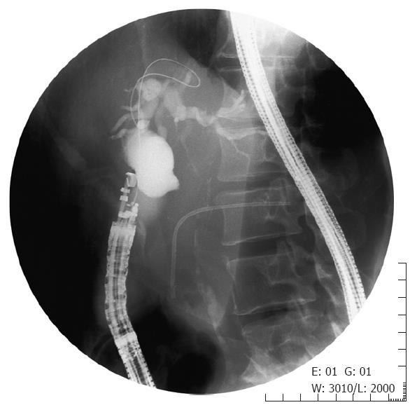
Figure 2 After EUS-guided puncture, contrast medium is injected into the bile duct for cholangiogram, a 0.
035-inch guidewire was inserted into the outer sheath (patient 3).
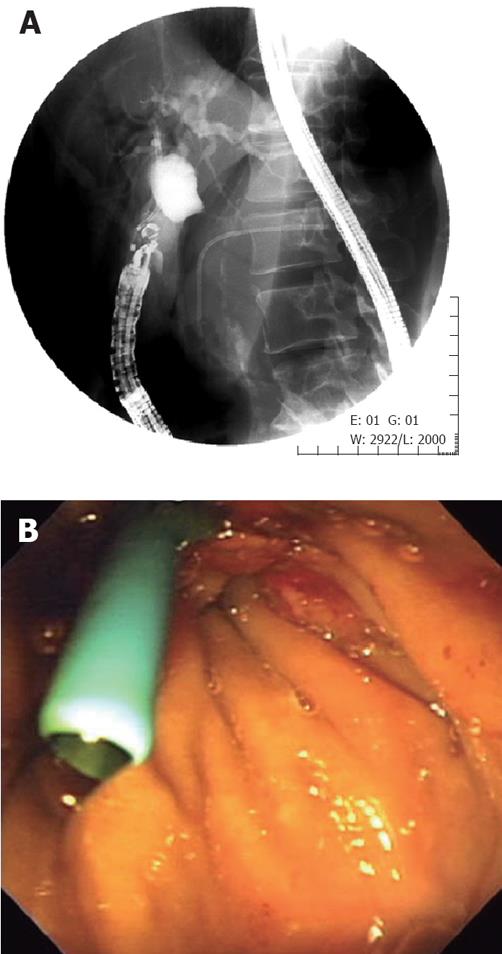
Figure 3 A: Choledochoduodenostomy was accomplished with a 7-Fr plastic stent in the apex of the duodenal bulb (patient 3); B: The stent was visible in the duodenal bulb (patient 3).
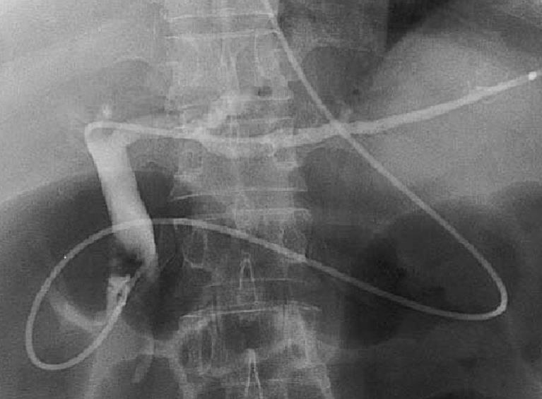
Figure 4 Choledochoduodenostomy was accomplished using a 5-Fr naso-biliary tube (patient 2).
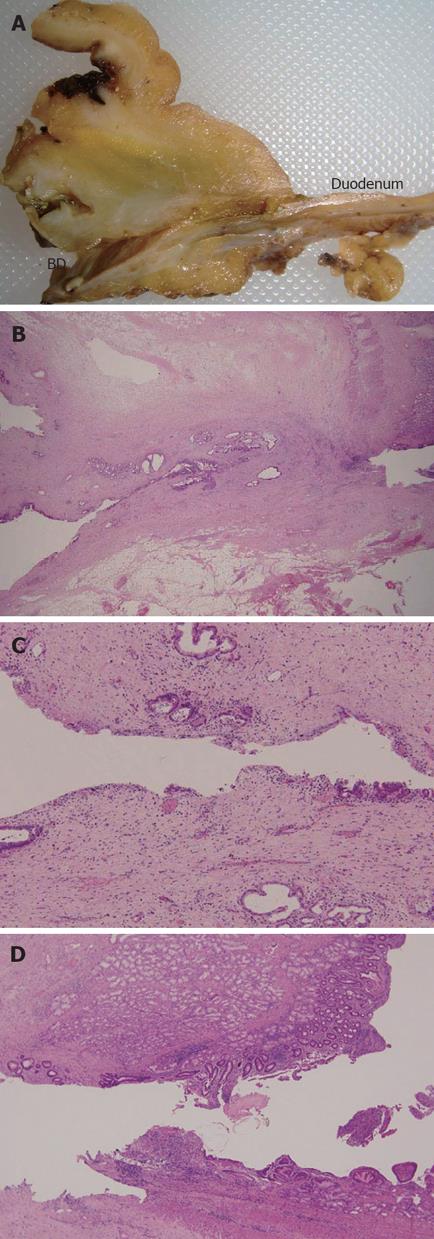
Figure 5 A: Macroscopic view of completed choledochoduodenostomy (patient 2); B: Surgical specimens showing the completed choledochoduodenostomy with mild inflammatory cell infiltrate adjacent to the sinus tract in the duodenal and bile duct walls (HE, × 20, patient 2); C: Magnification of bile duct site (HE, × 100, patient 2); D: Magnification of bile duct site (HE, × 100, patient 2).
BD: Bile duct.
- Citation: Itoi T, Itokawa F, Sofuni A, Kurihara T, Tsuchiya T, Ishii K, Tsuji S, Ikeuchi N, Moriyasu F. Endoscopic ultrasound-guided choledochoduodenostomy in patients with failed endoscopic retrograde cholangiopancreatography. World J Gastroenterol 2008; 14(39): 6078-6082
- URL: https://www.wjgnet.com/1007-9327/full/v14/i39/6078.htm
- DOI: https://dx.doi.org/10.3748/wjg.14.6078


