Copyright
©2008 The WJG Press and Baishideng.
World J Gastroenterol. Oct 14, 2008; 14(38): 5930-5932
Published online Oct 14, 2008. doi: 10.3748/wjg.14.5930
Published online Oct 14, 2008. doi: 10.3748/wjg.14.5930
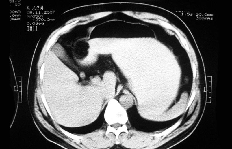
Figure 1 CT scan image showing a well-defined oval mass on the antro-pyloric part of stomach.
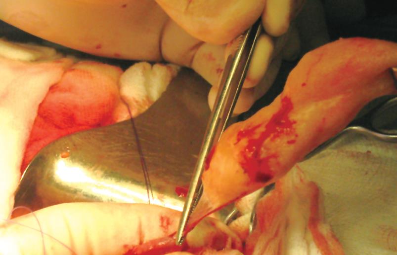
Figure 2 Intraoperative view showing a yellowish subserosal neoplasm on the anterior wall of gastric antrum during extirpation.
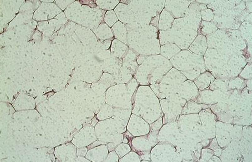
Figure 3 Histopathology confirming the diagnosis of lipoma containing mature fat cells with slight variation in size and shape (HE, × 100).
- Citation: Krasniqi AS, Hoxha FT, Bicaj BX, Hashani SI, Hasimja SM, Kelmendi SM, Gashi-Luci LH. Symptomatic subserosal gastric lipoma successfully treated with enucleation. World J Gastroenterol 2008; 14(38): 5930-5932
- URL: https://www.wjgnet.com/1007-9327/full/v14/i38/5930.htm
- DOI: https://dx.doi.org/10.3748/wjg.14.5930


