Copyright
©2008 The WJG Press and Baishideng.
World J Gastroenterol. Sep 14, 2008; 14(34): 5254-5260
Published online Sep 14, 2008. doi: 10.3748/wjg.14.5254
Published online Sep 14, 2008. doi: 10.3748/wjg.14.5254
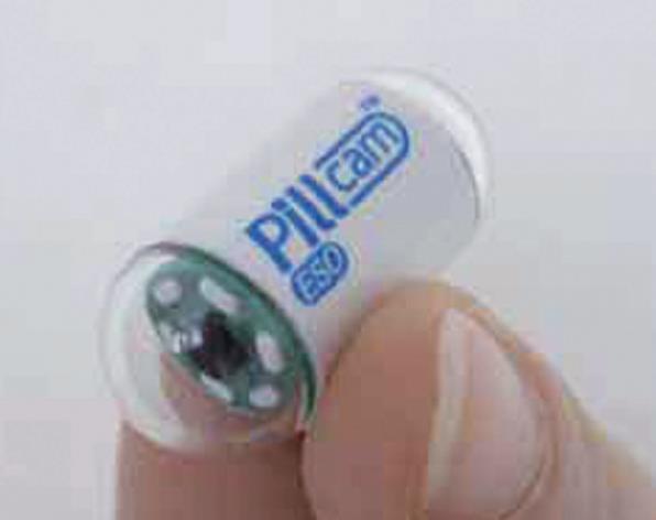
Figure 1 PillCam™ ESO.
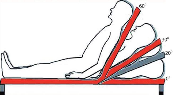
Figure 2 Ingestion protocol.
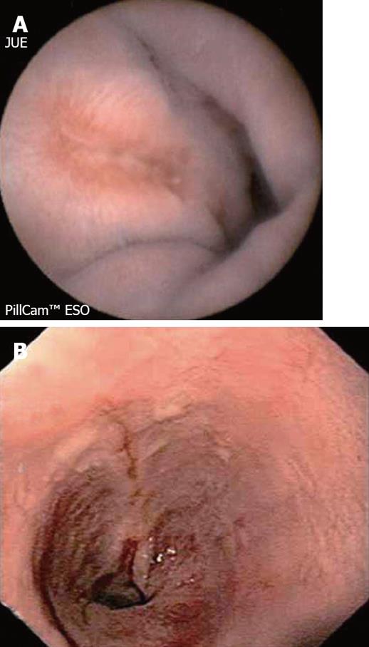
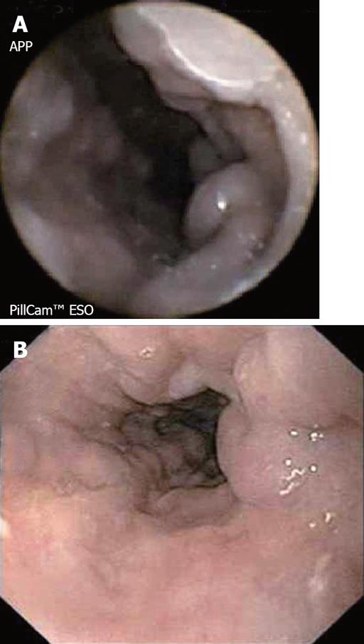
Figure 3 A: PillCam™ ESO image of erosive esophagitis; B: Upper endoscopy image of distal esophagus in the same patient.
Figure 4 A: PillCam™ ESO image of short segment Barrett´s esophagus; B: Subsequent upper endoscopy image which confirms capsule findings.
Figure 5 A: PillCam ESO™ image showing esophageal varices; B: Upper endoscopy image of distal esophagus in the same patient.
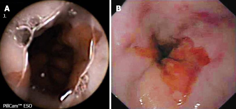
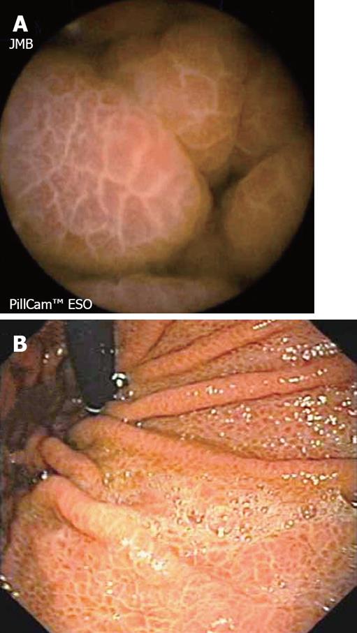
Figure 6 A: PillCam ESO™ image of the gastric wall showing portal hypertension gastropathy; B: Subsequent upper endoscopy image which confirms capsule findings.
- Citation: Fernandez-Urien I, Carretero C, Armendariz R, Muñoz-Navas M. Esophageal capsule endoscopy. World J Gastroenterol 2008; 14(34): 5254-5260
- URL: https://www.wjgnet.com/1007-9327/full/v14/i34/5254.htm
- DOI: https://dx.doi.org/10.3748/wjg.14.5254


