Copyright
©2008 The WJG Press and Baishideng.
World J Gastroenterol. Sep 14, 2008; 14(34): 5237-5244
Published online Sep 14, 2008. doi: 10.3748/wjg.14.5237
Published online Sep 14, 2008. doi: 10.3748/wjg.14.5237
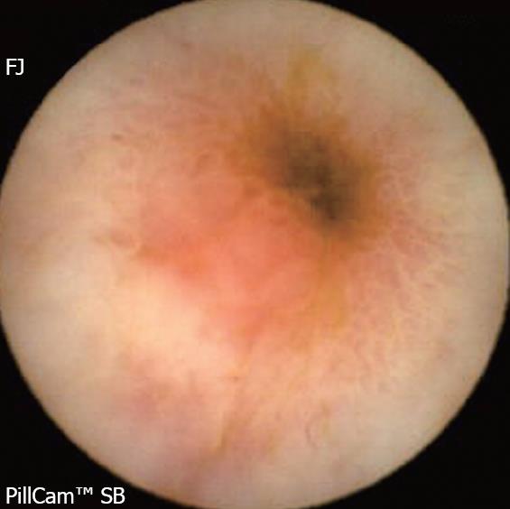
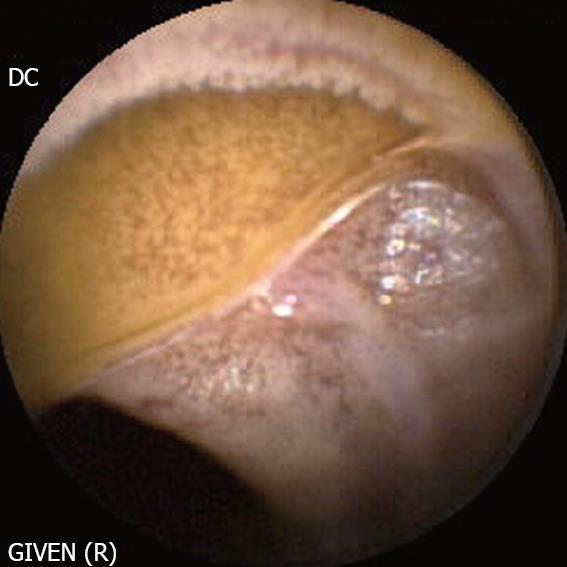
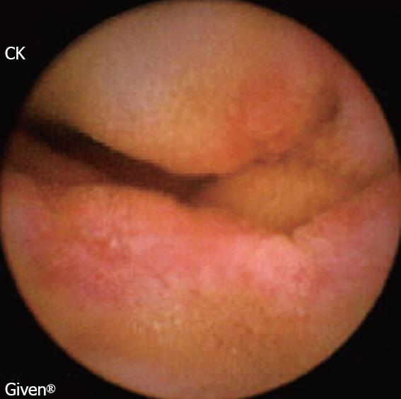
Figure 1 Intestinal stenosis caused by NSAIDs.
Capsule endoscopy shows narrowed segment, surrounded by an ulcerated but non-inflammatory mucosa.
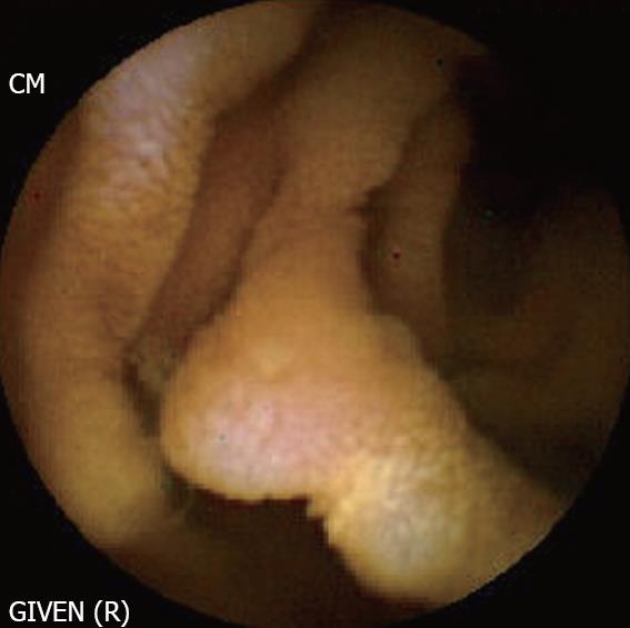
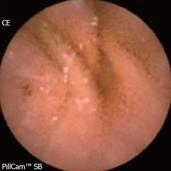
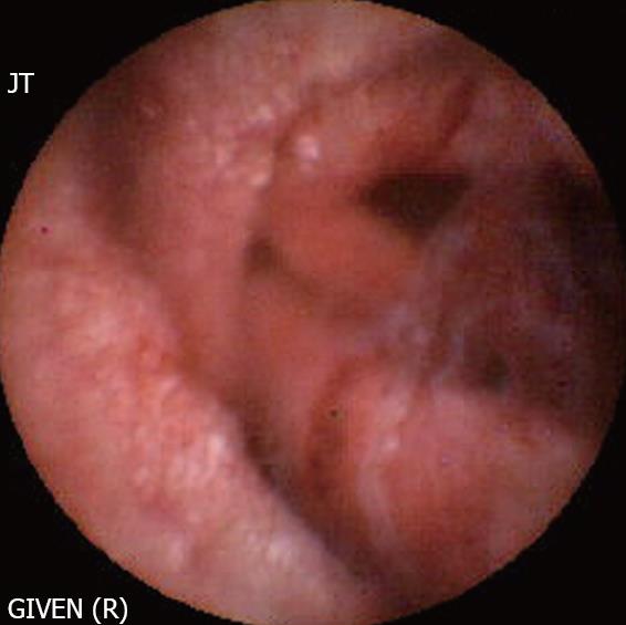
Figure 2 Waldmann’s disease.
The presence of dilated lymphatic vessels in the submucosa gives a whitish appearance to the mucosa. If lymphangiectasia becomes more prominent, it may protrude into the lumen.
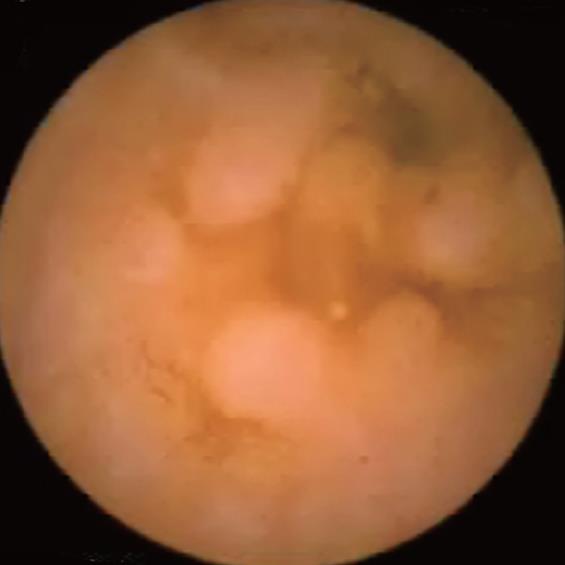
Figure 3 Common variable immunodeficiency disorder.
The characteristic lesions include polypoidal and nodular lesions of variable size, which are disseminated over the intestinal mucosa.
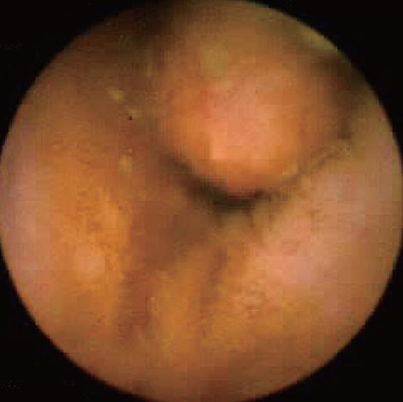
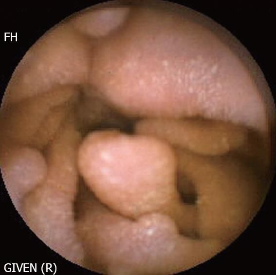
Figure 4 Peutz-Jeghers syndrome.
Presence of a large polyp in the ileum, in a young patient with Peutz-Jeghers syndrome. The polyp was ulcerated and required endoscopic resection.
Figure 5 Familial adenomatous polyposis.
Multiple polyps of regular shape are present in the ileum.
Figure 6 Acute Graft versus Host Disease (GVHD).
The lesions correspond to Stage III disease, manifesting as diffuse erythema and oedema of the mucosa, with a few erosions.
Figure 7 Hypobetalipoproteinemia.
The endoscopic picture is characterized by a diffuse whitish pattern of the mucosa due to the accumulation of fat vesicles in the enterocytes. The size of the intestinal villi is normal.
Figure 8 Churg and Strauss disease.
A large ulcer is present in the ileum.
Figure 9 Cytomegalovirus infection in a heart transplant patient.
The disease is characterized by the presence of bleeding ulcers and erosions.
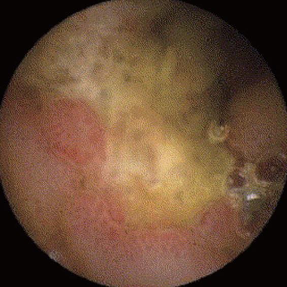
Figure 10 Whipple’s disease.
The endoscopic picture shows an oedematous and friable mucosa with erosions and serpiginous ulcers. The lesions may involve the entire length of the small bowel.
- Citation: Gay G, Delvaux M, Frederic M. Capsule endoscopy in non-steroidal anti-inflammatory drugs-enteropathy and miscellaneous, rare intestinal diseases. World J Gastroenterol 2008; 14(34): 5237-5244
- URL: https://www.wjgnet.com/1007-9327/full/v14/i34/5237.htm
- DOI: https://dx.doi.org/10.3748/wjg.14.5237


