Copyright
©2008 The WJG Press and Baishideng.
World J Gastroenterol. Aug 28, 2008; 14(32): 5096-5097
Published online Aug 28, 2008. doi: 10.3748/wjg.14.5096
Published online Aug 28, 2008. doi: 10.3748/wjg.14.5096
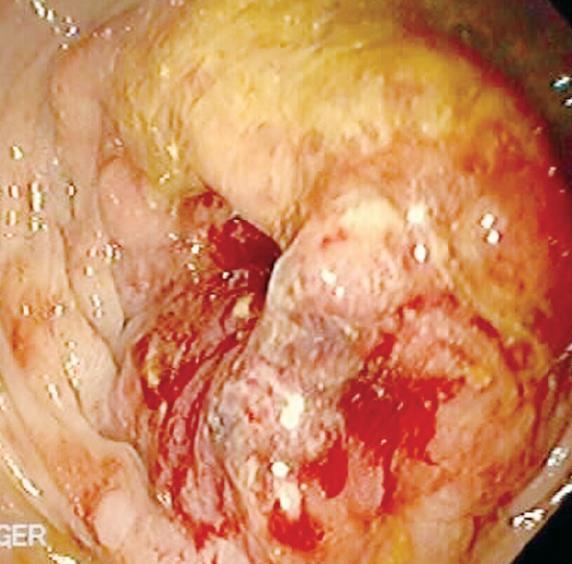
Figure 1 Colonoscopic view of an obstructing rectal cancer.
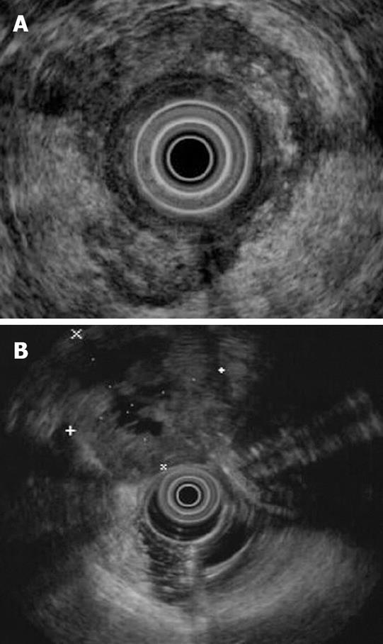
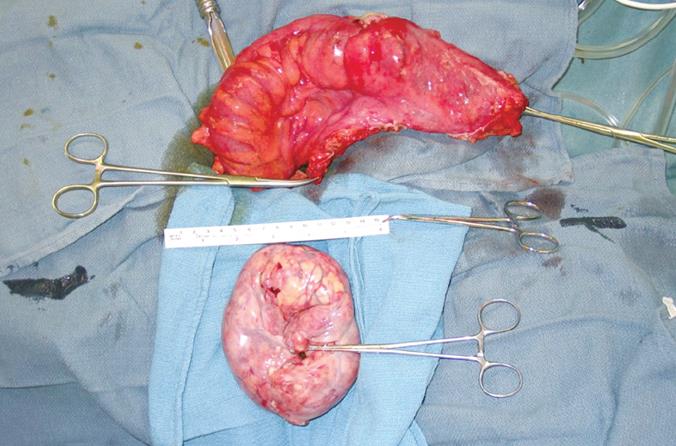
Figure 3 Gross pathologic findings at surgery showing the resected rectal carcinoma and the solitary ovarian metastases correlating with the findings in Figure 2.
- Citation: Moparty B, Gomez G, Bhutani MS. Large solitary ovarian metastasis from colorectal cancer diagnosed by endoscopic ultrasound. World J Gastroenterol 2008; 14(32): 5096-5097
- URL: https://www.wjgnet.com/1007-9327/full/v14/i32/5096.htm
- DOI: https://dx.doi.org/10.3748/wjg.14.5096


