Copyright
©2008 The WJG Press and Baishideng.
World J Gastroenterol. Jan 21, 2008; 14(3): 469-473
Published online Jan 21, 2008. doi: 10.3748/wjg.14.469
Published online Jan 21, 2008. doi: 10.3748/wjg.14.469
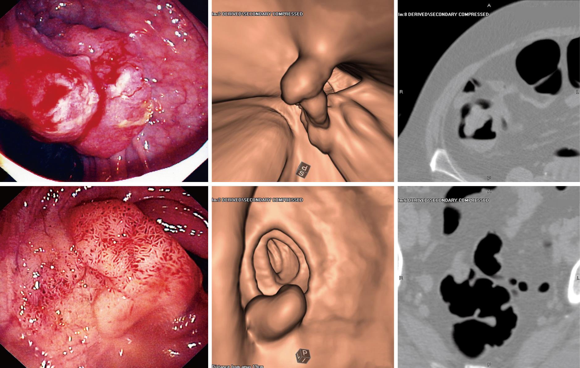
Figure 1 The figure shows the endoscopic appearance and images at CT colonography (3-D and 2-D) for 2 colonic lesions.
The upper panel shows a cancer in the ascending colon while the lower panel shows a large tubulovillous adenoma at the rectosigmoid junction.
- Citation: Roberts-Thomson IC, Tucker GR, Hewett PJ, Cheung P, Sebben RA, Khoo EW, Marker JD, Clapton WK. Single-center study comparing computed tomography colonography with conventional colonoscopy. World J Gastroenterol 2008; 14(3): 469-473
- URL: https://www.wjgnet.com/1007-9327/full/v14/i3/469.htm
- DOI: https://dx.doi.org/10.3748/wjg.14.469


