Copyright
©2008 The WJG Press and Baishideng.
World J Gastroenterol. Jan 21, 2008; 14(3): 441-447
Published online Jan 21, 2008. doi: 10.3748/wjg.14.441
Published online Jan 21, 2008. doi: 10.3748/wjg.14.441
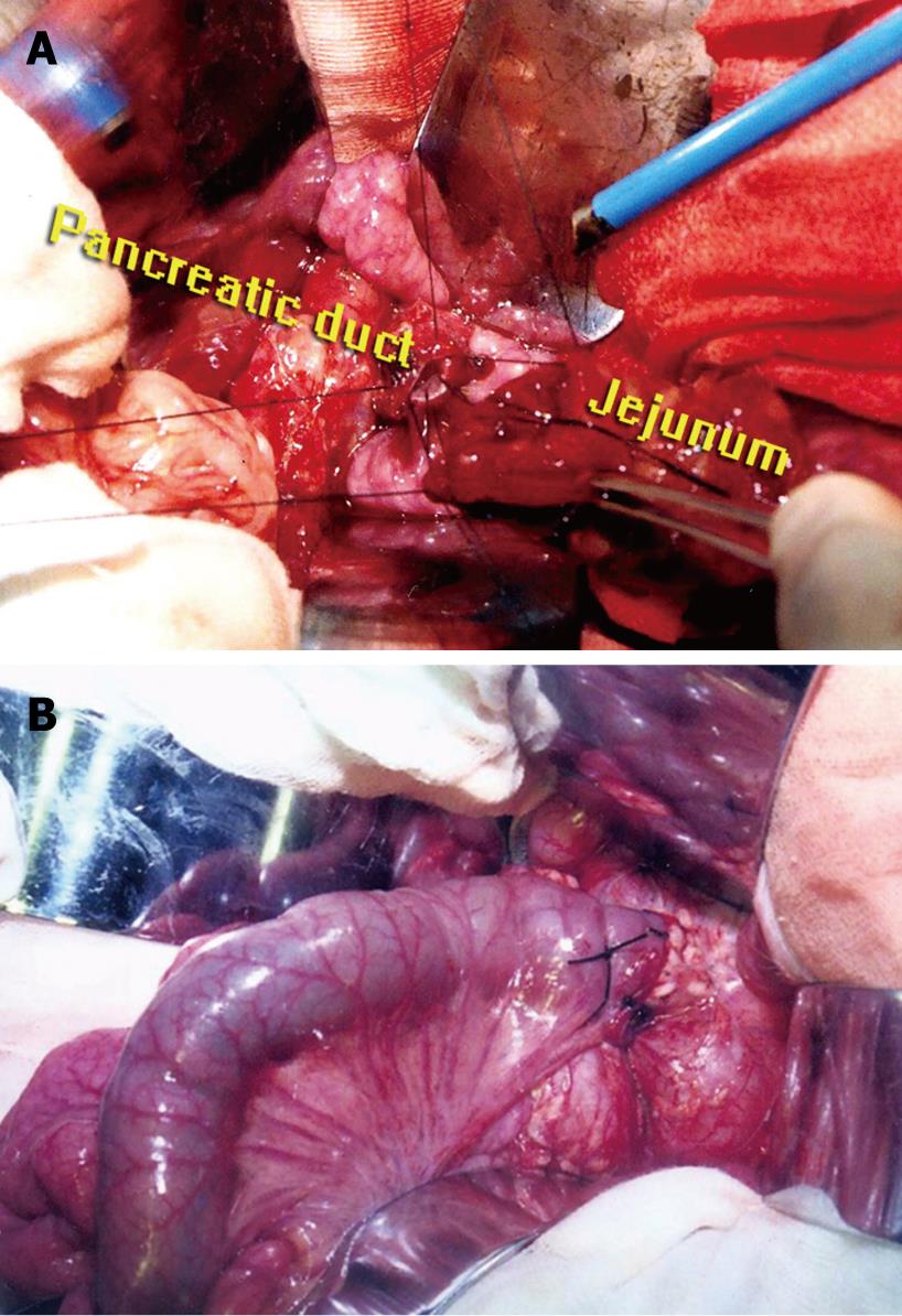
Figure 1 A: Pancreatic duct and jejunal mucosa are sutured, and stitch is only inserted to the submucosal layer of the jejunal wall; B: A piece of unabsorbable thread (7/0 silk) is used to bind together circumferentially the jejunal seromuscular sheath and pancreatic remnant.
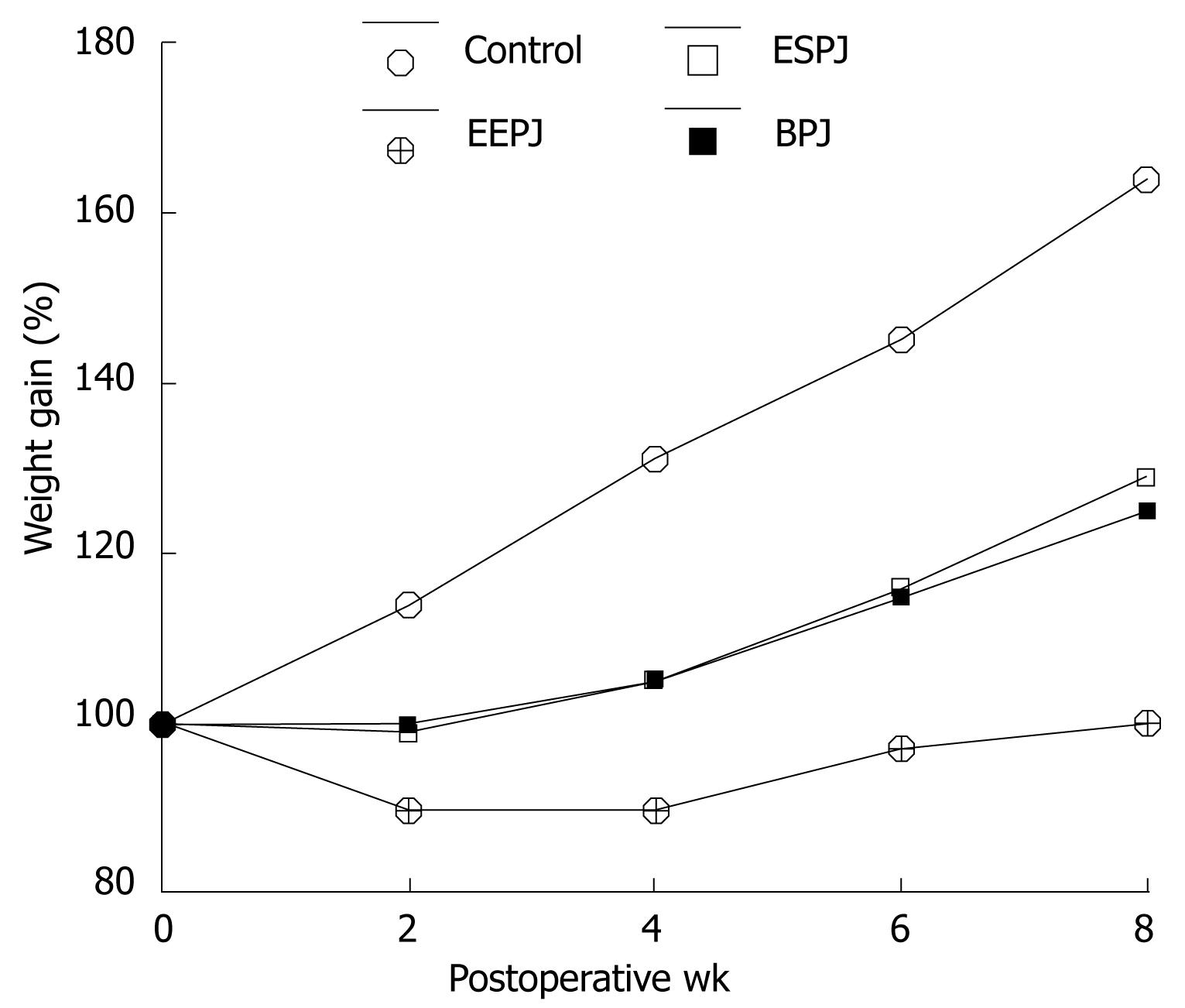
Figure 2 Percentage weight gain in pigs undergoing three types of pancreatoenteric anastomosis and in control pigs.
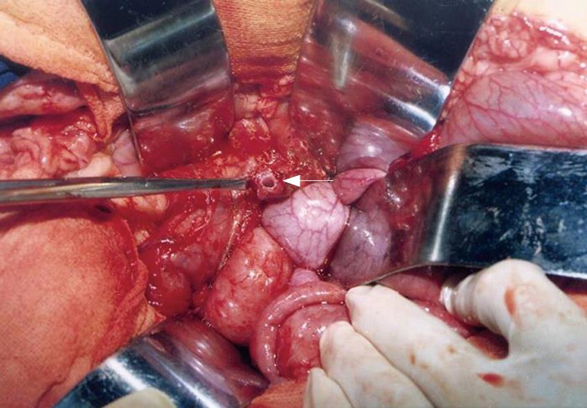
Figure 3 Duct at the cut surface of pancreatic stump was markedly dilated at the end of POW 4 after ligation of the main pancreatic duct.
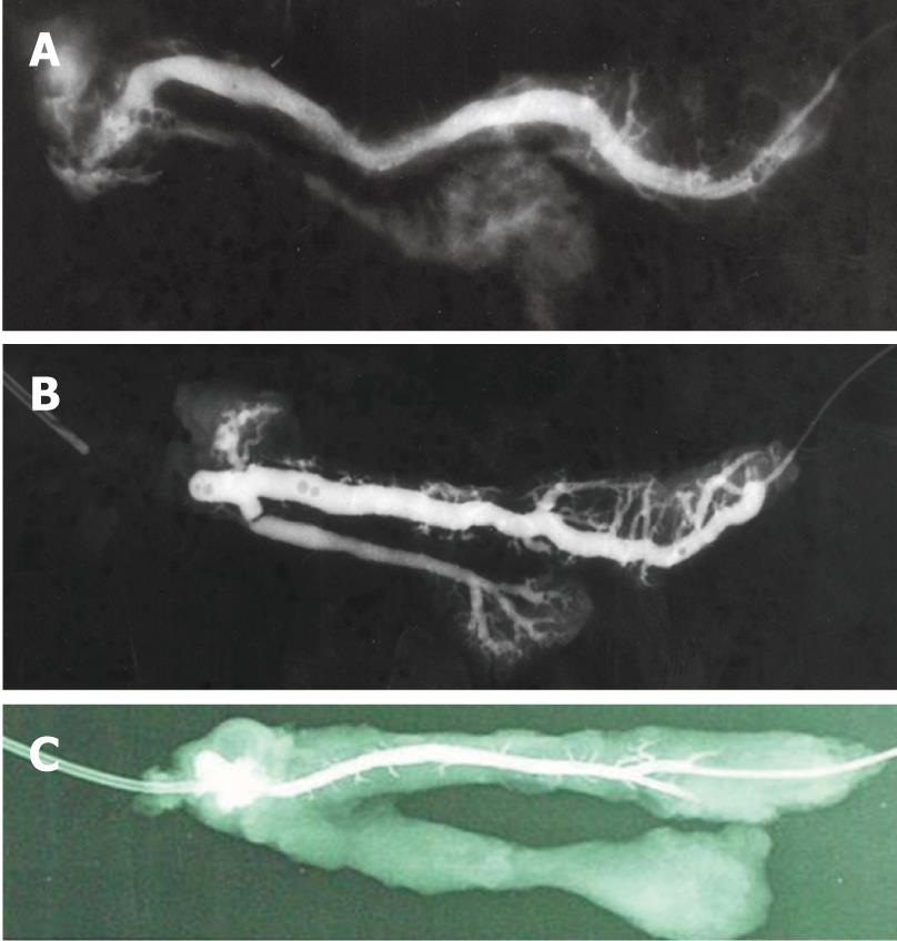
Figure 4 A: Anastomotic site was incompletely obstructed; B: Duct was blocked in the anastomotic region; C: Normal ductal patency without stenosis at the anastomotic site and without ductal dilatation proximal to it.
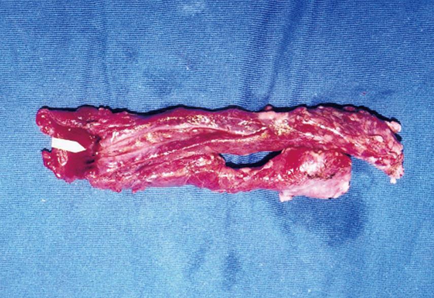
Figure 5 Longitudinal specimen reveals good connective tissue union between the pancreatic duct and the jejunal lumen.
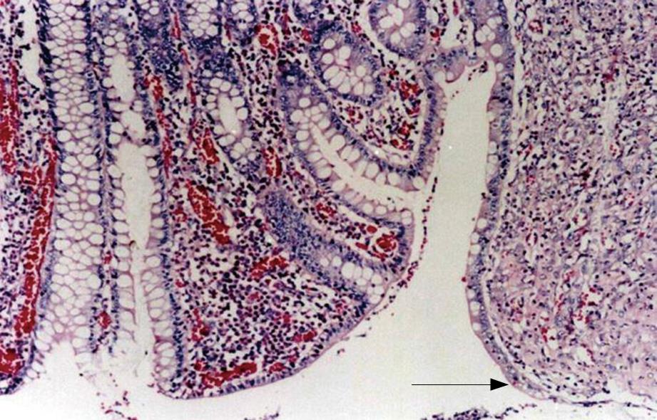
Figure 6 Section showing that the intestinal mucosa had fused with that of the pancreatic duct, with gradual and continuous changes from one to the other.
Original magnification × 100.
- Citation: Bai MD, Rong LQ, Wang LC, Xu H, Fan RF, Wang P, Chen XP, Shi LB, Peng SY. Experimental study on operative methods of pancreaticojejunostomy with reference to anastomotic patency and postoperative pancreatic exocrine function. World J Gastroenterol 2008; 14(3): 441-447
- URL: https://www.wjgnet.com/1007-9327/full/v14/i3/441.htm
- DOI: https://dx.doi.org/10.3748/wjg.14.441


