Copyright
©2008 The WJG Press and Baishideng.
World J Gastroenterol. Jan 21, 2008; 14(3): 421-427
Published online Jan 21, 2008. doi: 10.3748/wjg.14.421
Published online Jan 21, 2008. doi: 10.3748/wjg.14.421
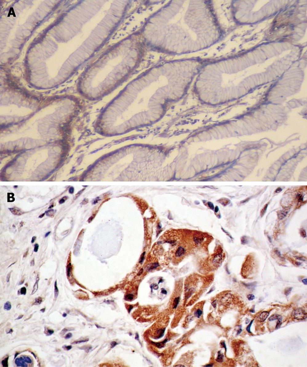
Figure 1 In situ hybridization of integrin β3 mRNA.
A: Negative expression in the plasma of non-tumor gastric mucosa (ISH, × 100); B: Positive expression in the plasma of gastric adenocarcinoma (ISH, × 200).
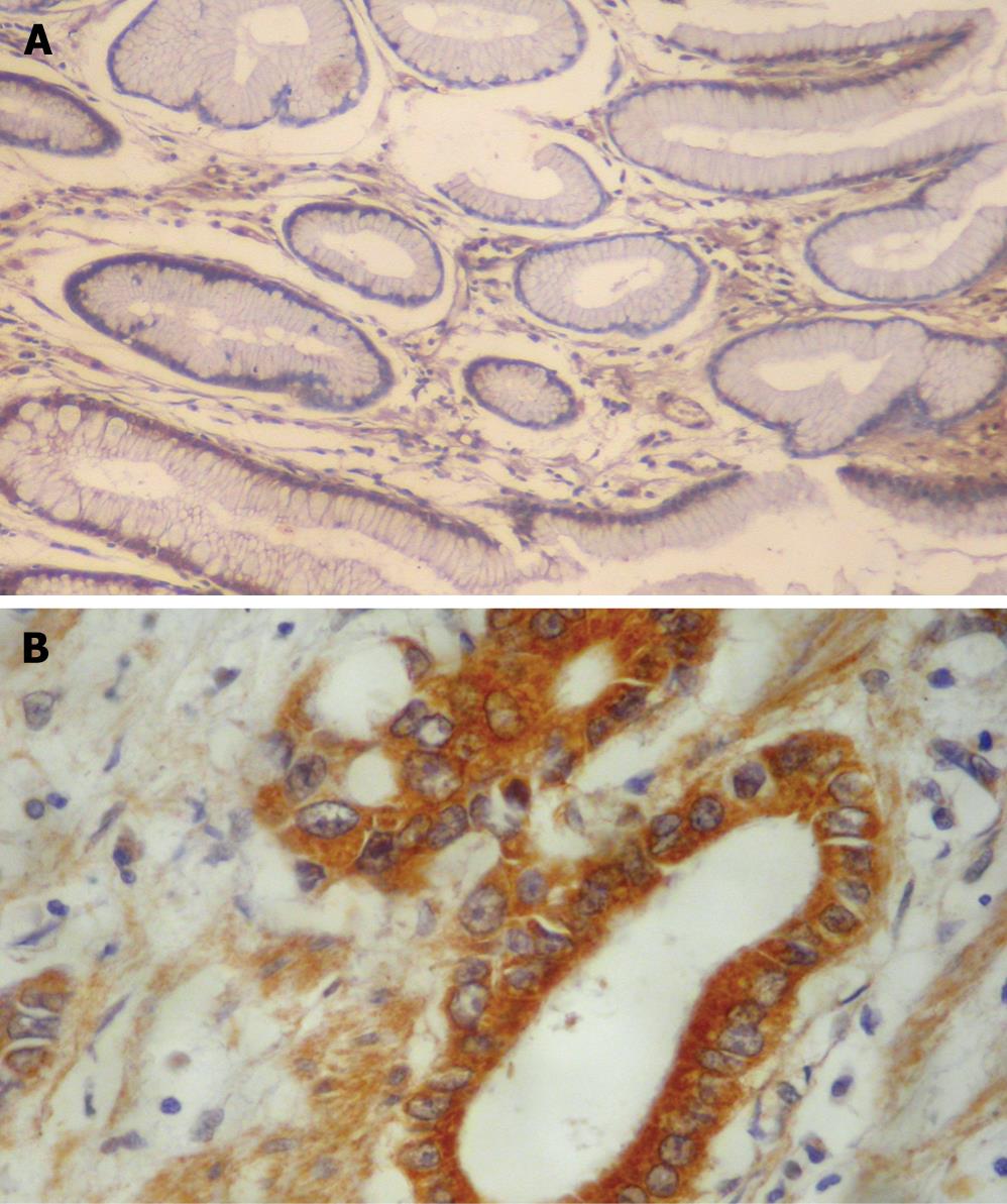
Figure 2 Immunohistochemical staining of VEGF.
A: Negative staining in the cytoplasm of non-tumor gastric mucosa (EnVision, × 100); B: Positive staining in the cytoplasm of gastric adenocarcinoma with greater omentum infiltration (EnVision, × 200).
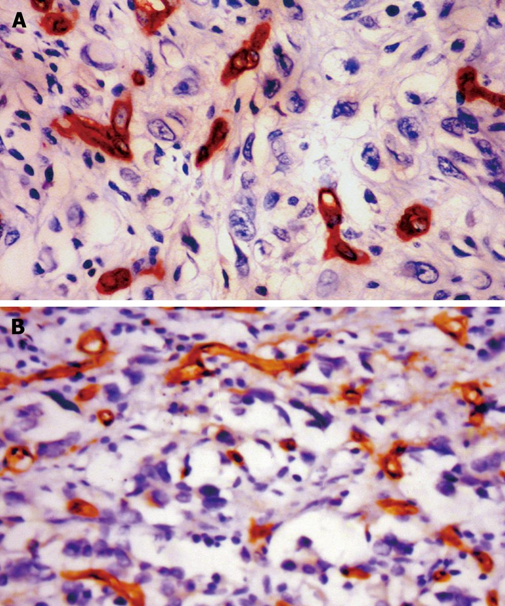
Figure 3 Microvessels in gastric cancer.
A: Positive staining for CD34 in the vascular endothelial cells of gastric adenocarcinoma with MVD < 54.9/mm2 (EnVision, × 400); B: Positive staining for CD34 in the vascular endothelial cells of gastric adenocarcinoma with MVD ≥ 54.9/mm2 (EnVision, × 400).
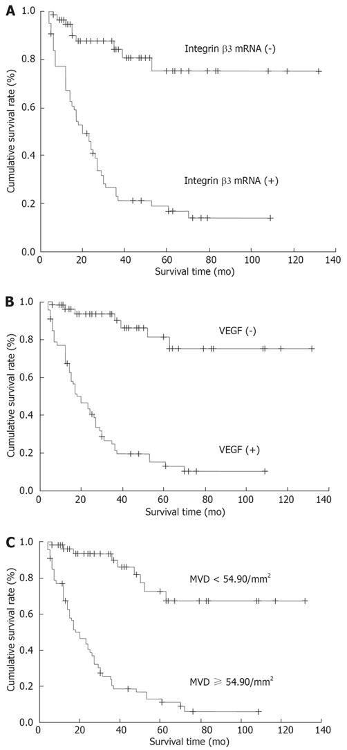
Figure 4 Kaplan-Meier survival curves.
A: Survival curves with positive and negative integrin β3 mRNA expression in gastric adenocarcinoma (P < 0.05); B: Survival curves with positive and negative VEGF expression in gastric adenocarcinoma (P < 0.05); C: Survival curves with MVD ≥ 54.90/mm2 and < 54.90/mm2 in gastric adenocarcinoma (P < 0.01).
- Citation: Li SG, Ye ZY, Zhao ZS, Tao HQ, Wang YY, Niu CY. Correlation of integrin β3 mRNA and vascular endothelial growth factor protein expression profiles with the clinicopathological features and prognosis of gastric carcinoma. World J Gastroenterol 2008; 14(3): 421-427
- URL: https://www.wjgnet.com/1007-9327/full/v14/i3/421.htm
- DOI: https://dx.doi.org/10.3748/wjg.14.421


