Copyright
©2008 The WJG Press and Baishideng.
World J Gastroenterol. Aug 7, 2008; 14(29): 4705-4708
Published online Aug 7, 2008. doi: 10.3748/wjg.14.4705
Published online Aug 7, 2008. doi: 10.3748/wjg.14.4705
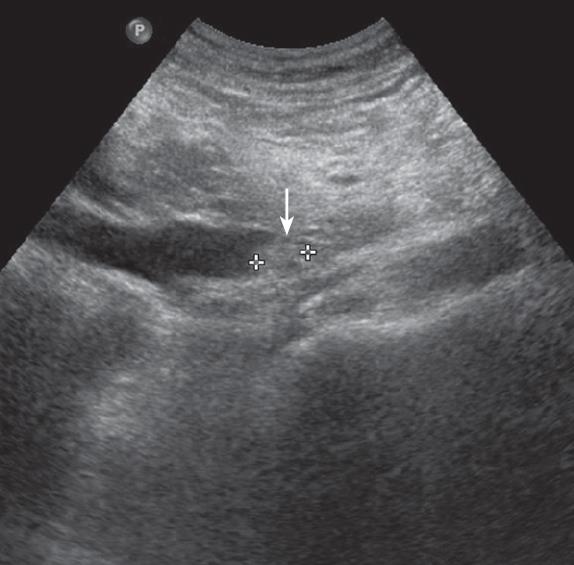
Figure 1 Ultrasound examination reveals a 2 cm sized non-shadowing mass and a dilated common bile duct (arrow).
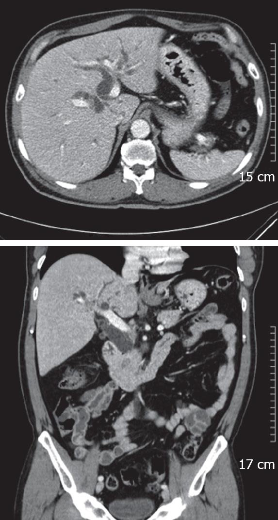
Figure 2 Computed tomography shows a diffuse dilatation of the common bile duct with an intraluminal mass in the distal common bile duct.
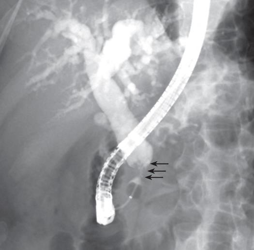
Figure 3 Endoscopic retrograde cholangiopancreatography shows a 2 cm round lobulated filling defect in the distal common bile duct (arrows).
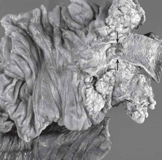
Figure 4 A photograph of the pylorus-preserving pancreaticoduodenectomy specimen shows a polypoid mass in the distal common bile duct, 2 cm x 1.
5 cm x 1.5 cm size (arrows).
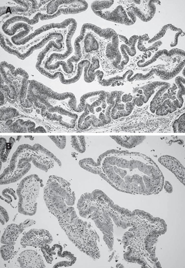
Figure 5 Microscopic features of the tubulovillous adenoma.
Hematoxylineosin stain. A: The elongated and pseoudostratification of nuclei are stained (× 40); B: Higher magnification on this slide demonstrates carcinoma in situ (asterisk; × 100).
-
Citation: Kim BS, Joo SH, Joo KR. Carcinoma
in situ arising in a tubulovillous adenoma of the distal common bile duct: A case report. World J Gastroenterol 2008; 14(29): 4705-4708 - URL: https://www.wjgnet.com/1007-9327/full/v14/i29/4705.htm
- DOI: https://dx.doi.org/10.3748/wjg.14.4705


