Copyright
©2008 The WJG Press and Baishideng.
World J Gastroenterol. Aug 7, 2008; 14(29): 4701-4704
Published online Aug 7, 2008. doi: 10.3748/wjg.14.4701
Published online Aug 7, 2008. doi: 10.3748/wjg.14.4701
Figure 1 Endoscopic image showing a bleeding ulcer in the second part of the duodenum (A), the endoscopic clipping procedure revealing no other bleeding site detected by upper and lower gastrointestinal endoscopy (B), and follow-up gastrointestinal endoscopic view 12 d after hemostatic procedure (C).
Only one hemoclip remained on the scar of the duodenal ulcer.
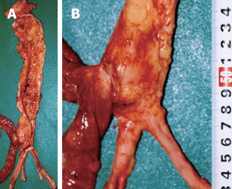
Figure 2 The presence of a firm adhesion between the AAA and the digestive duodenal wall located on the third part of duodenum (A), and magnification of the firm adhesion between the anterior site of the abdominal aorta and the posterior serosa of the duodenum (B).
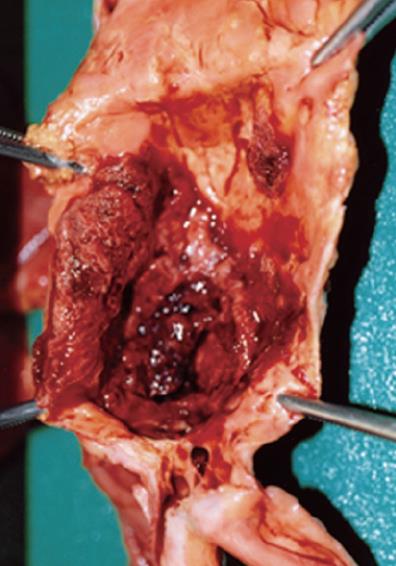
Figure 3 The AAA has severe atherosclerosis with calcification and clotted blood on the aneurysm inside the aortic lumen.
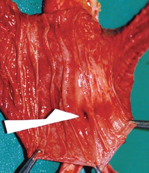
Figure 4 A rupture and the size of a pinhole with no degeneration or inflammation of the third part of the duodenal mucosa as indicated by the white arrow.
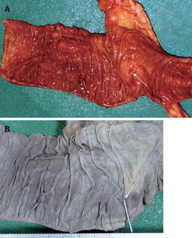
Figure 5 A scar on the clipped ulcer indicated by the black arrow, papilla Vater indicated by the black triangle, and a rupture on the third part of duodenal mucosa located in front of the firm adhesion indicated by the white arrow in the duodenal mucosa specimen (A), and a postclipping scar indicated by the black arrow and a rupture indicated by the white arrow in the formalin-fixed specimen of the duodenal mucosa (B).
The rupture was located approximately 6 cm from the anal side of the postclipping scar.
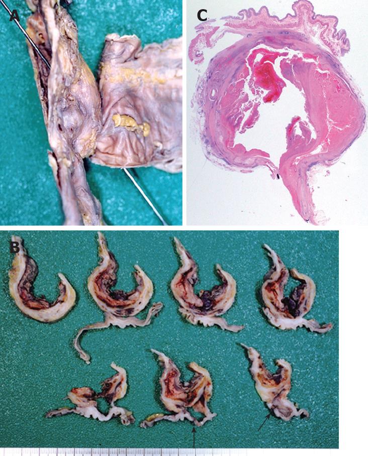
Figure 6 Formalin-fixed duodenum and aorta specimen demonstrating a sonde inserted from the rupture on the duodenal mucosa to the inside of the aortic lumen (A), horizontal cross sections showing a firm adhesion between the aorta and its wall as well as a rupture (black arrow) on the third part of the duodenum (B); and a microphotograph of a horizontal cross section showing an adhesion between the duodenum and the aorta with atherosclerosis-contained clotted blood (C).
The histology of the duodenal appears normal (HE, × 5).
- Citation: Ihama Y, Miyazaki T, Fuke C, Ihama Y, Matayoshi R, Kohatsu H, Kinjo F. An autopsy case of a primary aortoenteric fistula: A pitfall of the endoscopic diagnosis. World J Gastroenterol 2008; 14(29): 4701-4704
- URL: https://www.wjgnet.com/1007-9327/full/v14/i29/4701.htm
- DOI: https://dx.doi.org/10.3748/wjg.14.4701


