Copyright
©2008 The WJG Press and Baishideng.
World J Gastroenterol. Aug 7, 2008; 14(29): 4633-4642
Published online Aug 7, 2008. doi: 10.3748/wjg.14.4633
Published online Aug 7, 2008. doi: 10.3748/wjg.14.4633
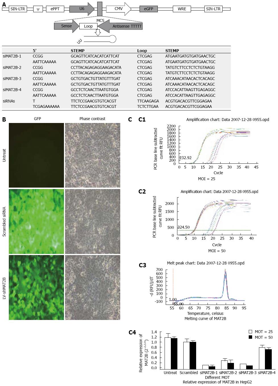
Figure 1 A: Each consist of a hairpin as was demonstrated; B: HepG2 cells were without treatment (untreated), and infected with control lentivirus (scrambled siRNA) or LV-shMAT2B.
The cells were infected (MOI = 25), GFP expression and the phase contrast images of the same areas were taken after 72 h. C: msRNA level of MAT2B after HepG2 cell was treated with LV-shMAT2B in different MOI (C1 and C2) detected by real-time PCR (C4), melting curve of MAT2 B (C3).
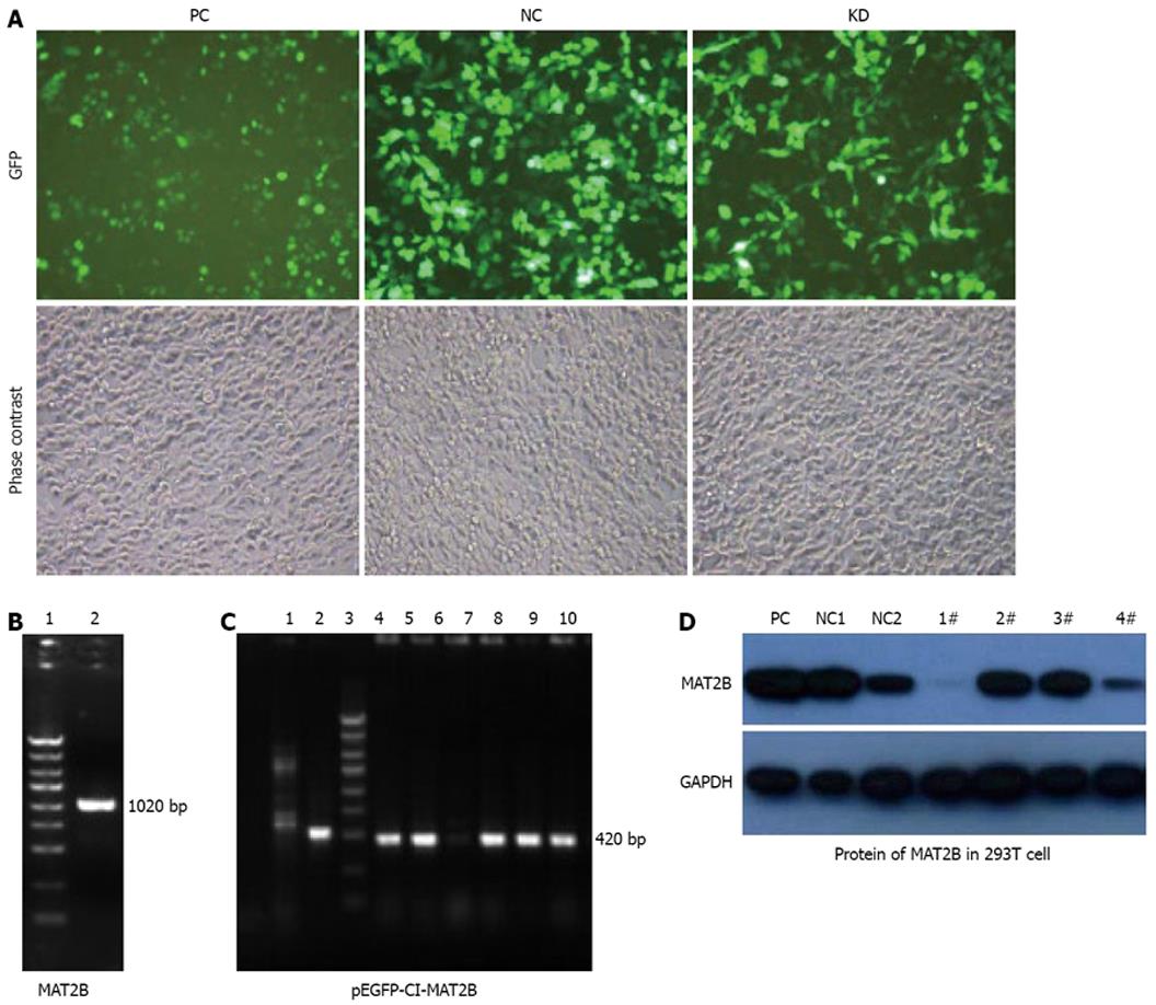
Figure 2 MAT2B gene was cloned from the cDNA library with production of 1020 bp by PCR (B), pEGFP-C1-MAT2B was constructed after restricted enzyme cutting and connection with production of 420 bp by PCR as showed in Figure2C.
Plasmids containing siMAT2B (pGCL-GFP-siMAT2B) were co-transfected with pEGFP-C1-MAT2B in 293T cell, phase contrast and GFP expression under a fluorescent microscope was taken after 72 h (A). Protein level of MAT2B in 293T cells was detected by western blot (D). pGCL-GFP-siMAT2B-1 and -4 can significantly knock down expression of MAT2B at the protein level (D). PC, pcDNA3.1; NC, non-silence control vector. Lanes 1-4: pGCL-GFP-siMAT2B-1, -2, -3 and -4, respectively.
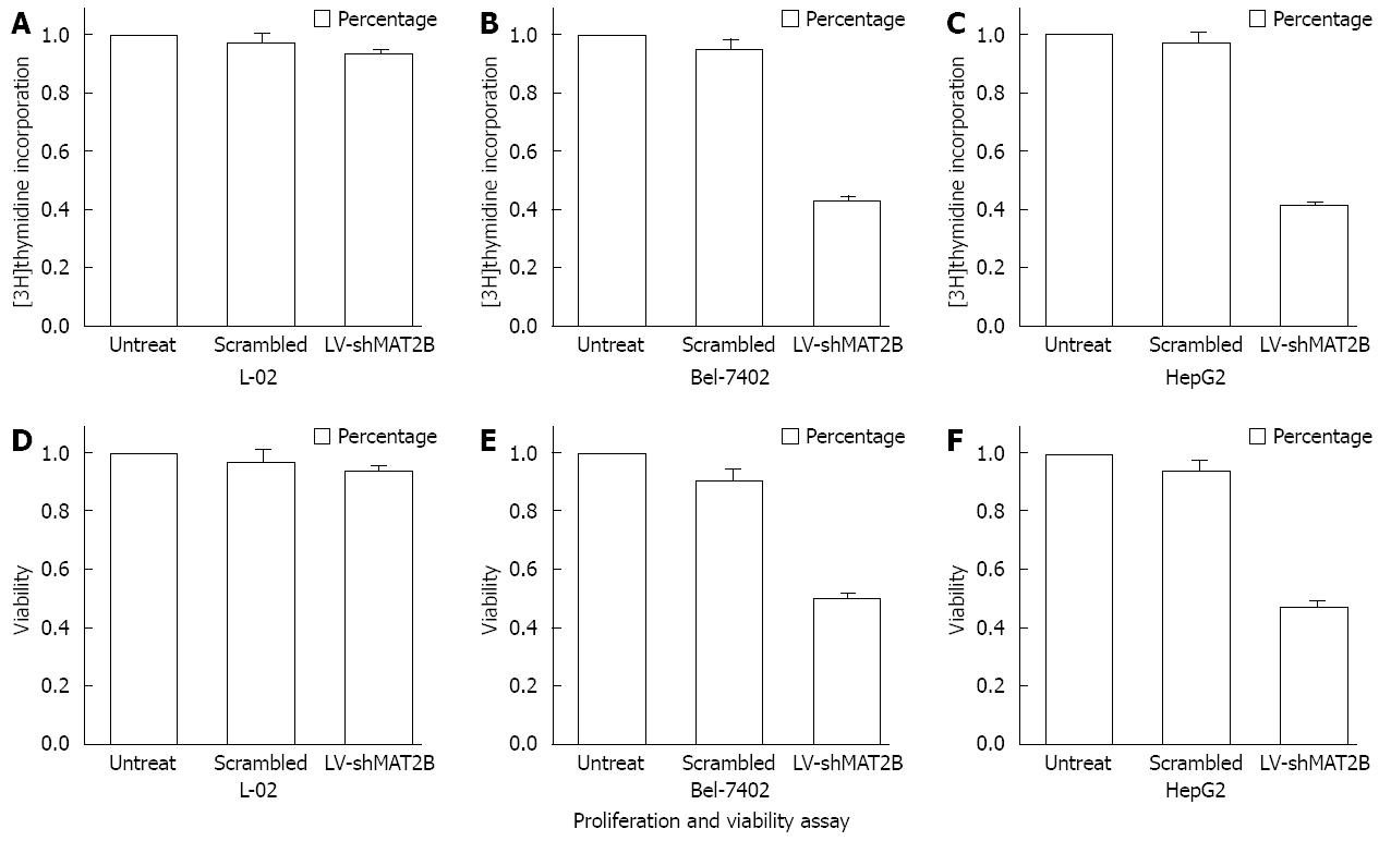
Figure 3 Proliferation of L-02 cell and HCC cells Bel-7402, HepG2 were shown in Figure 3A-C.
The relative value of [3H]thymidine incorporation was measured after cells were treated with LV-shMAT2B. Percentage cell viability of L-02 cells and HCC cells Bel-7402. HepG2 were counted with MTT after cells were treated with LV-shMAT2B Figure 2D-F. It was seen that LV-shMAT2B can inhibit cell viability and proliferation of HCC cells but had no effect on normal liver cells.
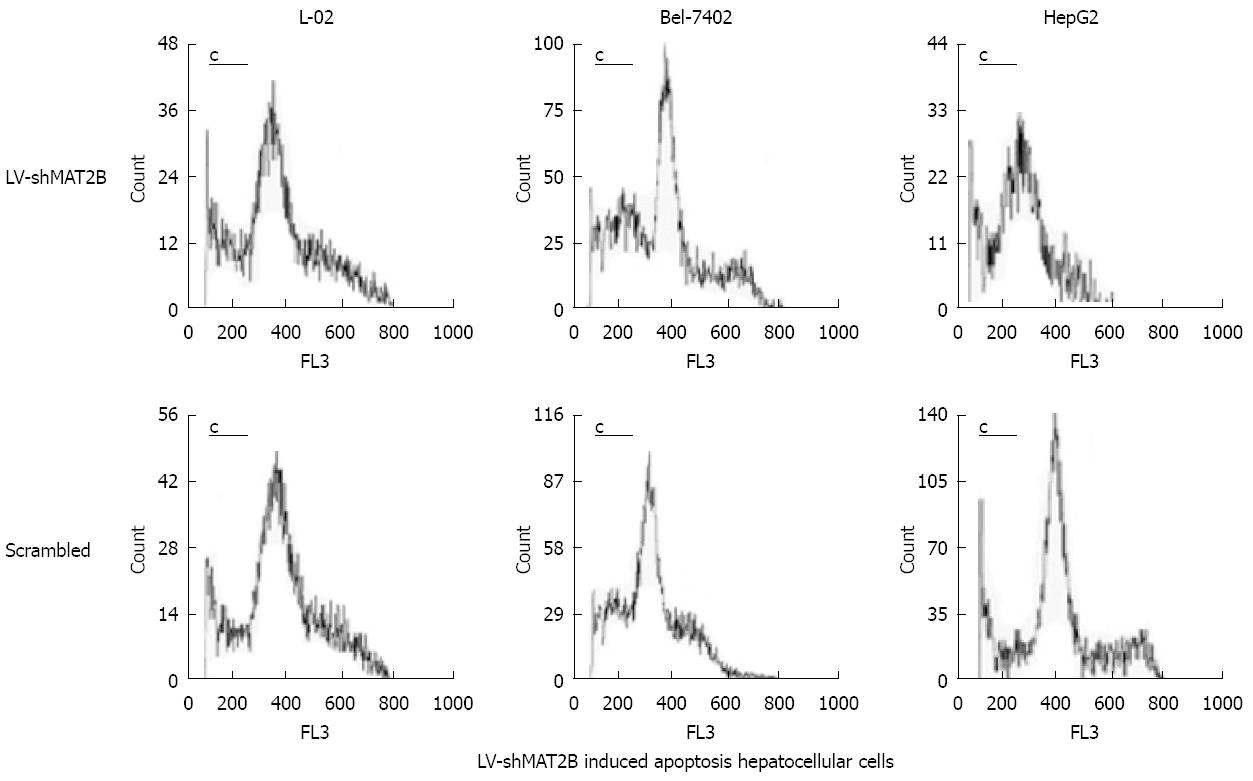
Figure 4 Cell apoptosis caused by LV-shMAT2B was increased in human hepatocellular cancer cells Bel-7402 and HepG2 compared to controls induced by siRNAs, but they all had no impact on the normal hepatic cell (L-02) which does not express MAT2B.
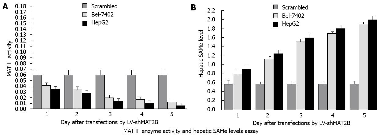
Figure 5 MAT II enzyme activity was gradually decreased from 1 d to 5 d in a time-dependent manner after transfected with LV-shMAT2B, but remained relatively unchanged in cells treated with siRNAs.
In human cancer cells Bel-7402 and HepG2 (A), Hepatic SAM level in the two HCC cells were increased by LV-shMAT2B in a time-dependent manner, but remained relatively unchanged in cells treated with siRNAs (B).
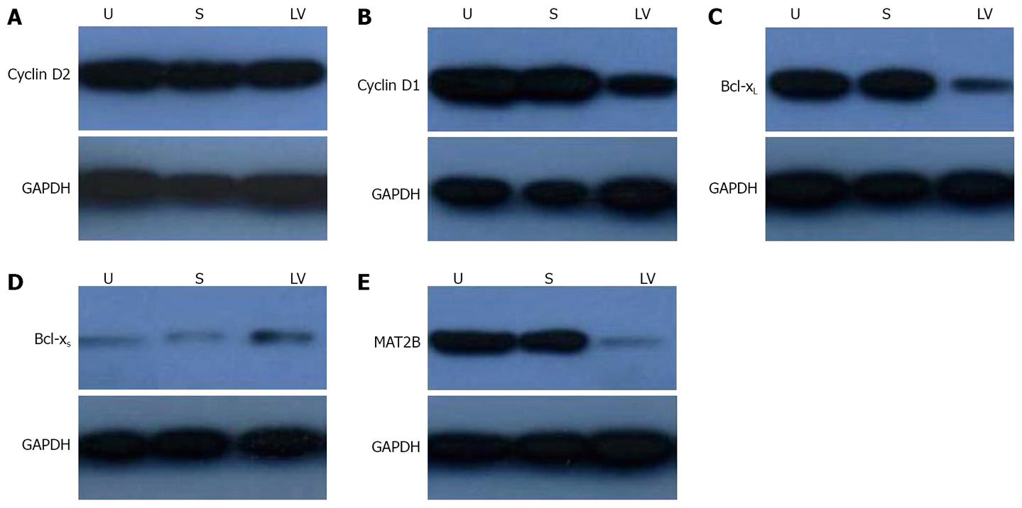
Figure 6 After HepG2 cells were transfected with LV-shMAT2B for 72 h, western blot was used to detect the protein levels of cyclin D1 and cyclin D2, bcl-xL and bcl-xs,
results show the expressions of cyclin D1 (B)and Bcl-xL (C) were decreased compared to controls and untreated cells.
The expression of bcl-xs (D) was increased but
that of cyclin D2 did not change (A). The expression of MAT2B was suppressed by LV-shMAT2B significantly (E). U, untreated HepG2 cell; S, HepG2 cell treated with
scrambled siRNA; LV, HepG2 cells treated with LV-shMAT2B. The protein level of MAT2B was measured by western blot.
- Citation: Wang Q, Liu QY, Liu ZS, Qian Q, Sun Q, Pan DY. Lentivirus mediated shRNA interference targeting MAT2B induces growth-inhibition and apoptosis in hepatocellular carcinoma. World J Gastroenterol 2008; 14(29): 4633-4642
- URL: https://www.wjgnet.com/1007-9327/full/v14/i29/4633.htm
- DOI: https://dx.doi.org/10.3748/wjg.14.4633


