Copyright
©2008 The WJG Press and Baishideng.
World J Gastroenterol. Jul 28, 2008; 14(28): 4583-4585
Published online Jul 28, 2008. doi: 10.3748/wjg.14.4583
Published online Jul 28, 2008. doi: 10.3748/wjg.14.4583
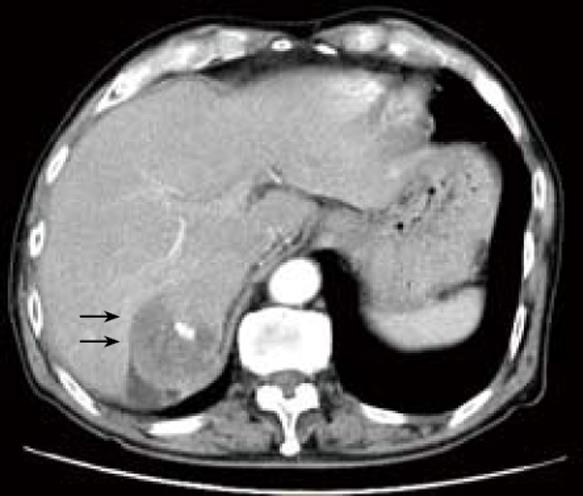
Figure 1 Computed tomography images showing a 7.
5-cm liver tumor (arrows) arising from the caudate lobe in case 1(A), which appears to invade the transverse colon directly (arrows) (B).
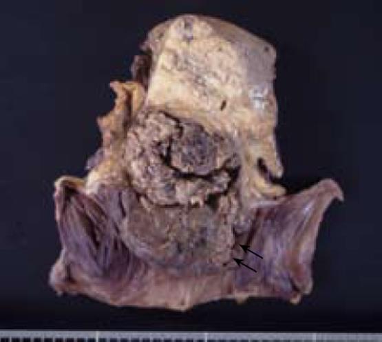
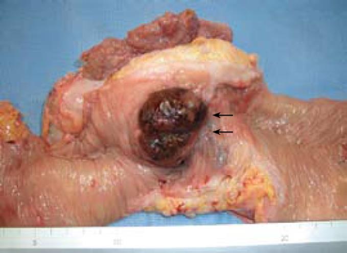
Figure 2 Macroscopic appearance of the surgical specimen in case 1.
The liver tumor invades the colon (arrows).
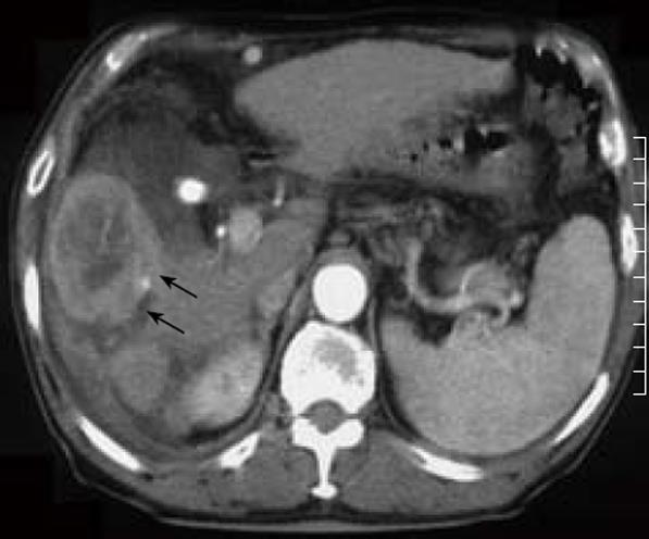
Figure 3 Computed tomography images showing a 6-cm liver tumor invades the colon and diaphragm (arrows) in case 2.
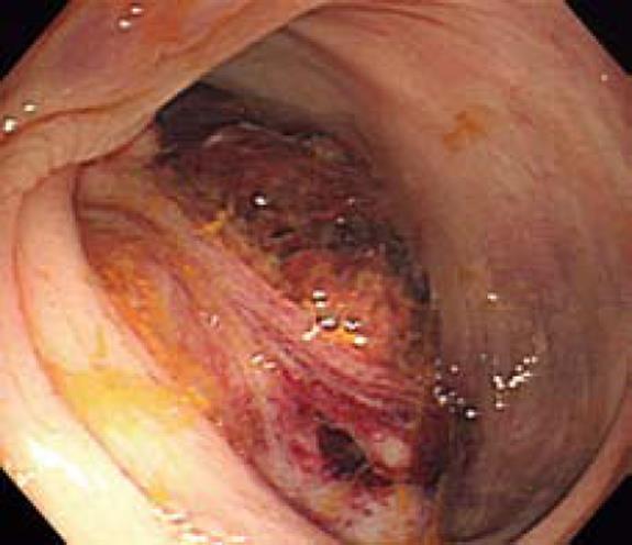
Figure 4 Colonoscopic view showing a hemorrhagic and lobulated tumor with a smooth surface is seen in the ascending colon in case 2.
Figure 5 Macroscopic appearance of the surgical specimen in case 2.
The liver tumor invades the colon (arrows).
- Citation: Hirashita T, Ohta M, Iwaki K, Kai S, Shibata K, Sasaki A, Nakashima K, Kitano S. Direct invasion to the colon by hepatocellular carcinoma: Report of two cases. World J Gastroenterol 2008; 14(28): 4583-4585
- URL: https://www.wjgnet.com/1007-9327/full/v14/i28/4583.htm
- DOI: https://dx.doi.org/10.3748/wjg.14.4583


