Copyright
©2008 The WJG Press and Baishideng.
World J Gastroenterol. Jul 28, 2008; 14(28): 4573-4575
Published online Jul 28, 2008. doi: 10.3748/wjg.14.4573
Published online Jul 28, 2008. doi: 10.3748/wjg.14.4573
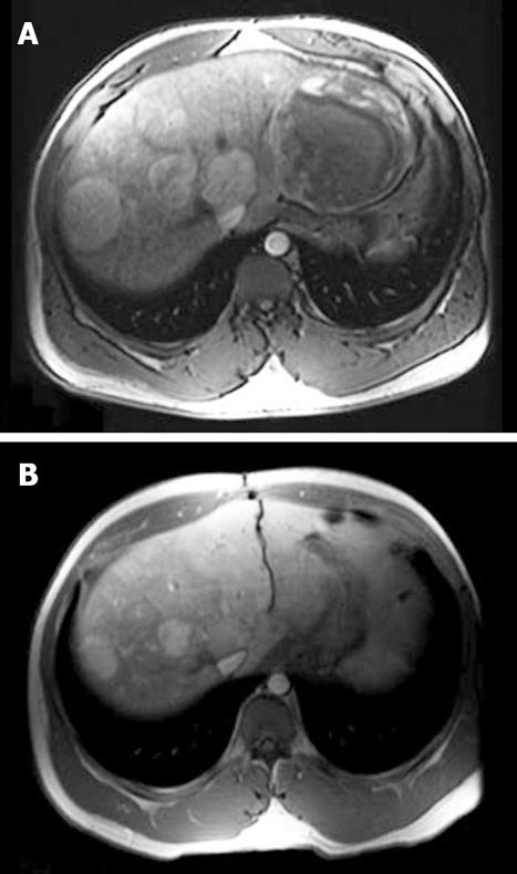
Figure 1 T1-weighted magnetic resonance imaging (MRI) of multiple hepatic adenomas.
A: MRI at initial presentation demonstrates a heterogeneous-appearing, well-circumscribed mass measuring 10.6 cm x 10.6 cm in segments 2 and 3 of the liver and several smaller masses in the right liver. The largest mass has an enhancing capsule and demonstrates areas of internal T1 hyperintensity and hypointensity, as well as T2 hyperintensity, characteristic of an adenoma with intralesional hemorrhage; B: Three months after left lateral segmentectomy and steroid cessation, the lesions in the right liver appear approximately 40% smaller and enhance more homogeneously on T1-weighted MRI, indicating regression. Images have been electronically adjusted to illustrate lesions.
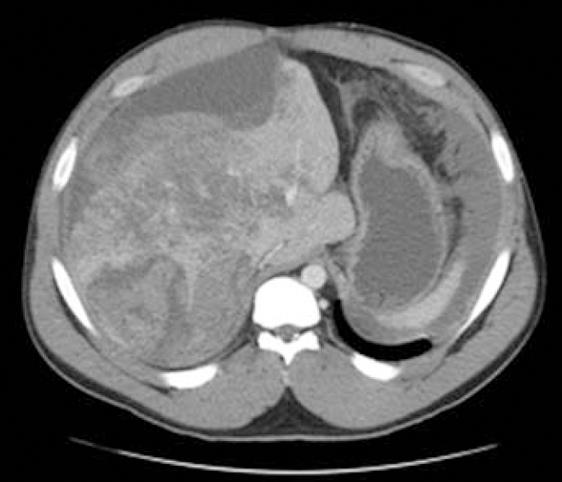
Figure 2 Abdominal CT at second presentation with abdominal pain after resumption of steroid abuse.
A heterogeneous-appearing, right hepatic mass measuring 7.2 cm × 7.7 cm and a large subcapsular hematoma are seen, indicating that one of the hepatic adenomas has enlarged since the previous presentation and has hemorrhaged spontaneously. Image has been electronically altered.
- Citation: Martin NM, Dayyeh BKA, Chung RT. Anabolic steroid abuse causing recurrent hepatic adenomas and hemorrhage. World J Gastroenterol 2008; 14(28): 4573-4575
- URL: https://www.wjgnet.com/1007-9327/full/v14/i28/4573.htm
- DOI: https://dx.doi.org/10.3748/wjg.14.4573


