Copyright
©2008 The WJG Press and Baishideng.
World J Gastroenterol. Jul 28, 2008; 14(28): 4540-4545
Published online Jul 28, 2008. doi: 10.3748/wjg.14.4540
Published online Jul 28, 2008. doi: 10.3748/wjg.14.4540
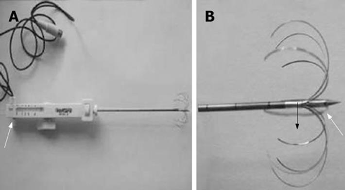
Figure 1 A: The white arrow indicates the injection hole; B: The white arrow indicates the side hole, and the black arrow indicates hooked array radiofrequency needles
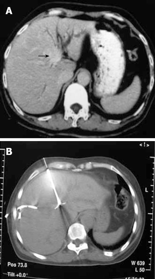
Figure 2 A case with type IIIb Klatskin tumor.
A: Plain CT scan reveals that the tumor is in the main trunk of left hepatic duct with dilatation of branches; B: The patient underwent RFA under CT guidance after internal and external drainage for two weeks.
Figure 3 A case with type IV Klatskin tumor.
A: The plain CT reveals that the tumor is in the portal hepatic region with dilatation of the left and right hepatic ducts; B: The patient underwent RFA under CT guidance after internal and external drainage for 2 wk; C: The plain CT scan reveals a liquidized and necrotic region of the tumor in the portal hepatic region without contrast enhancement 2 mo after RFA.
- Citation: Fan WJ, Wu PH, Zhang L, Huang JH, Zhang FJ, Gu YK, Zhao M, Huang XL, Guo CY. Radiofrequency ablation as a treatment for hilar cholangiocarcinoma. World J Gastroenterol 2008; 14(28): 4540-4545
- URL: https://www.wjgnet.com/1007-9327/full/v14/i28/4540.htm
- DOI: https://dx.doi.org/10.3748/wjg.14.4540


