Copyright
©2008 The WJG Press and Baishideng.
World J Gastroenterol. Jul 28, 2008; 14(28): 4529-4534
Published online Jul 28, 2008. doi: 10.3748/wjg.14.4529
Published online Jul 28, 2008. doi: 10.3748/wjg.14.4529
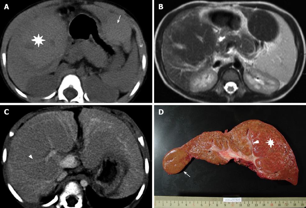
Figure 1 A 3-year-old BA girl with segments 5-8 (asterisk) and segment 2 (arrow) MRN.
A: The density of the MRN is slightly higher than that in the surrounding liver parenchyma during pre-enhanced phase of the CT; B: FSE/T2WI MRI shows a lower signal intensity in the MRN than in the surrounding liver; C: During portovenous phase of the CT, the tubular structure and splaying portal veins can be seen in the MRN (arrowhead); D: The explanted liver and intra-tumoral portal tract can be seen (arrowhead).
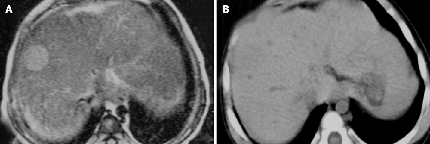
Figure 2 A 16-month-old BA girl with a 2.
1 cm MRN in segment 8. A: During pre-enhanced phase of the CT, no nodule can be seen; B: The signal intensity is hyperinstense on T2WI MRI.
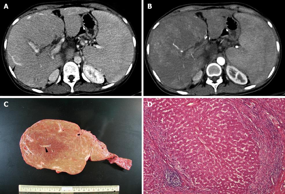
Figure 3 Patchy arterial enhancement within the mass in arterial phase of the CT (A), tubular structure (arrowhead) within the mass during portovenous phase of the CT (B), the explanted liver and intra-tumoral portal tract (arrowhead) (C), and microscopic examination (HE) showing uniform benign-looking liver cells arranged in one- to two-cell thick plates with intervening sinusoids and surrounding fibrous septa infiltrated with lymphocyte (D) in a 3-year-old BA girl with 13 cm MRN in right liver.
- Citation: Liang JL, Cheng YF, Concejero AM, Huang TL, Chen TY, Tsang LLC, Ou HY. Macro-regenerative nodules in biliary atresia: CT/MRI findings and their pathological relations. World J Gastroenterol 2008; 14(28): 4529-4534
- URL: https://www.wjgnet.com/1007-9327/full/v14/i28/4529.htm
- DOI: https://dx.doi.org/10.3748/wjg.14.4529


