Copyright
©2008 The WJG Press and Baishideng.
World J Gastroenterol. Jul 21, 2008; 14(27): 4400-4402
Published online Jul 21, 2008. doi: 10.3748/wjg.14.4400
Published online Jul 21, 2008. doi: 10.3748/wjg.14.4400
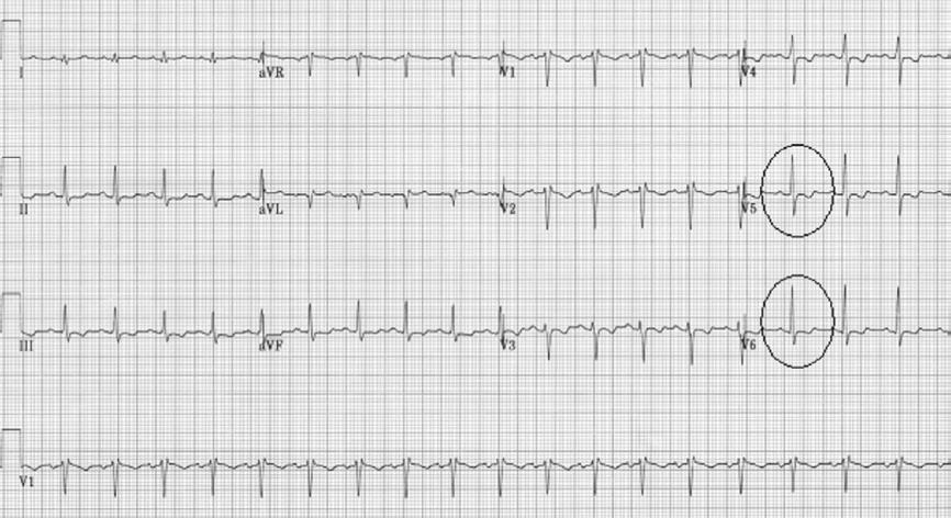
Figure 1 Non-specific ST-T wave changes with T-wave inversion in the lateral leads (circles).
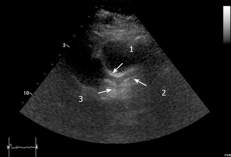
Figure 2 Echocardiogram-parasternal short-axis view showing mildly dilated left main coronary artery (1), left anterior descending artery (2), left circumflex coronary artery (3).
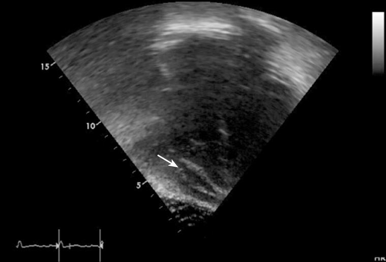
Figure 3 Echocardiogram-4-chamber view showing pericardial effusion (arrow).
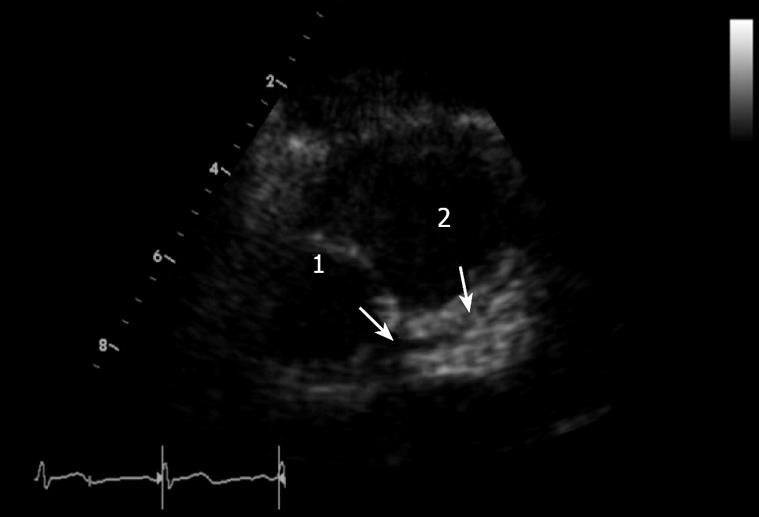
Figure 4 Echocardiogram-parasternal short-axis view showing normal left main coronary artery (1), normal left anterior descending artery (2).
- Citation: Atay O, Radhakrishnan K, Arruda J, Wyllie R. Severe chest pain in a pediatric ulcerative colitis patient after 5-aminosalicylic acid therapy. World J Gastroenterol 2008; 14(27): 4400-4402
- URL: https://www.wjgnet.com/1007-9327/full/v14/i27/4400.htm
- DOI: https://dx.doi.org/10.3748/wjg.14.4400


