Copyright
©2008 The WJG Press and Baishideng.
World J Gastroenterol. Jul 21, 2008; 14(27): 4395-4399
Published online Jul 21, 2008. doi: 10.3748/wjg.14.4395
Published online Jul 21, 2008. doi: 10.3748/wjg.14.4395
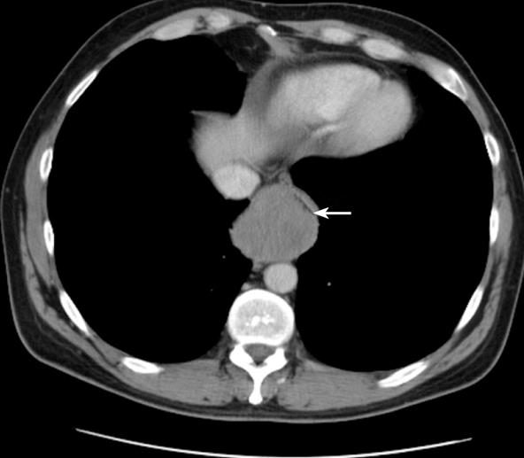
Figure 1 CT scan of chest highlighting the large distal esophageal mass.
Note the impressive constriction of the esophageal lumen (arrow).
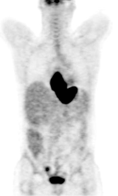
Figure 2 Whole body PET scan demonstrates intensely increased metabolic activity corresponding to the large esophagogastric mass.
There is also focal increased activity in a right iliac node.
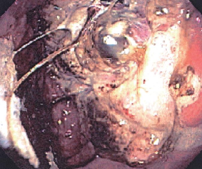
Figure 3 Upper Endoscopic evaluation shows esophageal mass with ulcerative features which extended into the gastric fundus.
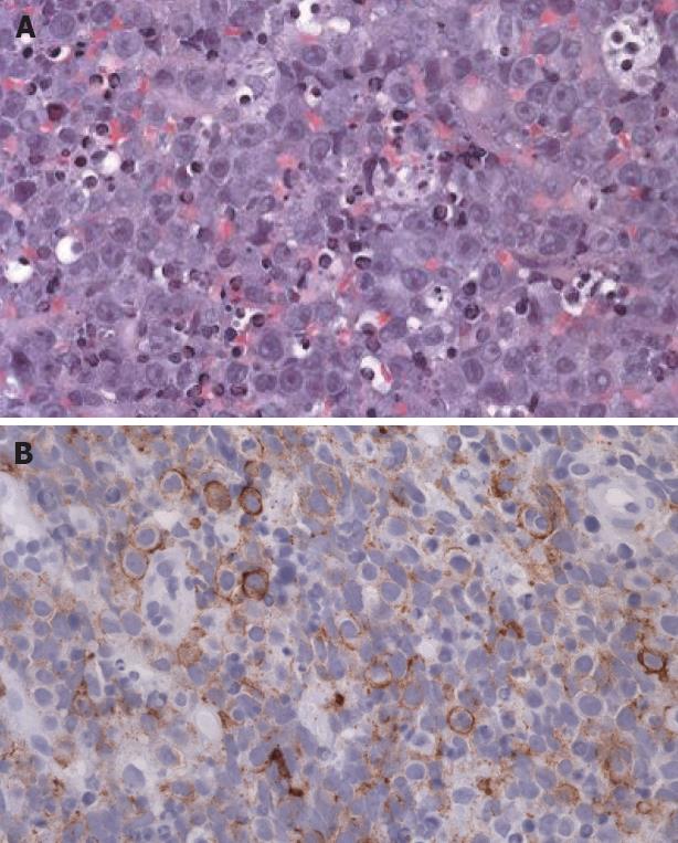
Figure 4 A: HE image shows a poorly differentiated, malignant neoplasm composed of irregular sheets of cells.
Cells are cytologically atypical with somewhat eccentric vesicular nuclei and prominent nucleoli (× 40); B: Immunohistochemistry shows patchy but strong staining of tumor cells for CD138 (× 40).
- Citation: Mani D, Jr DGG, Aboulafia DM. AIDS-associated plasmablastic lymphoma presenting as a poorly differentiated esophageal tumor: A diagnostic dilemma. World J Gastroenterol 2008; 14(27): 4395-4399
- URL: https://www.wjgnet.com/1007-9327/full/v14/i27/4395.htm
- DOI: https://dx.doi.org/10.3748/wjg.14.4395


