Copyright
©2008 The WJG Press and Baishideng.
World J Gastroenterol. Jul 14, 2008; 14(26): 4196-4203
Published online Jul 14, 2008. doi: 10.3748/wjg.14.4196
Published online Jul 14, 2008. doi: 10.3748/wjg.14.4196
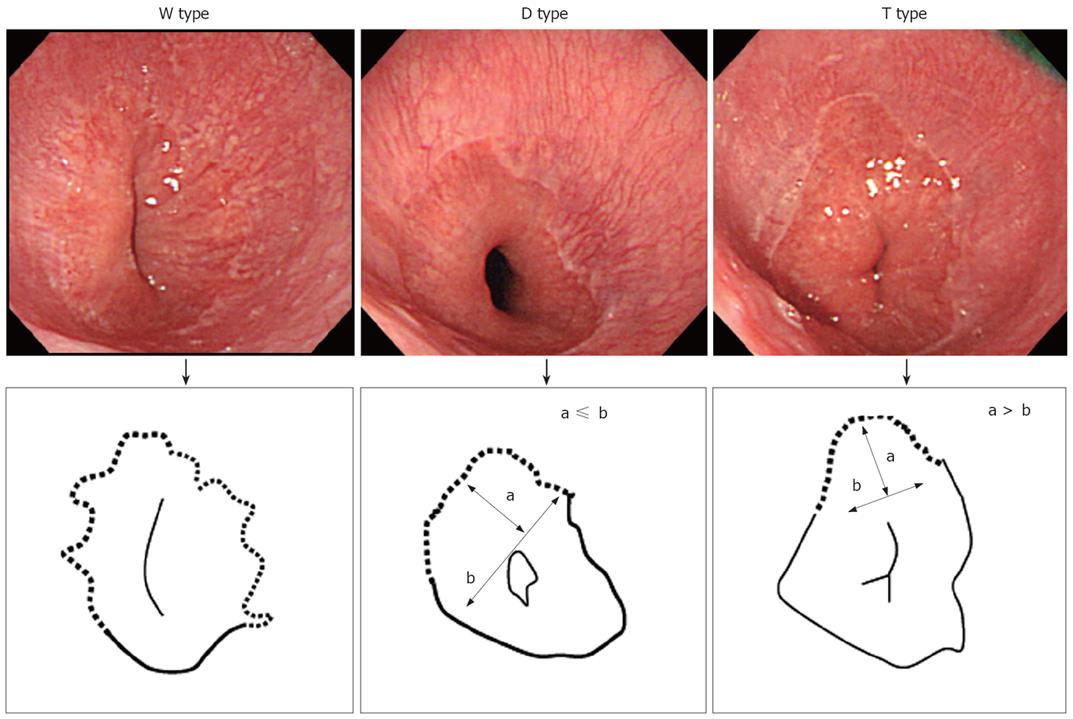
Figure 1 The shape of ESEM.
We originally divided ESEM into 3 types based on its shape. The W type, in which the ESEM was defined by a columnar epithelium which extended continuously from the gastric lumen to the esophagus, in which the uppermost extent of the visible red columnar epithelium could be observed as a wave-like formation (W). The broken line in the illustration shows the ESEM. The D type, in which the length of basal part of the ESEM (b) was longer than the length of the major part of the ESEM (a; i.e. a < b) The columnar epithelium of the ESEM is observed as a dome-like shape (D). The broken line in the illustration shows the ESEM. The T type in which the length of the major axis of the ESEM (a) is longer than the basal part of ESEM (b; i.e. a ≥ b). The columnar epithelium of the ESEM is observed as a tongue-like shape (T).
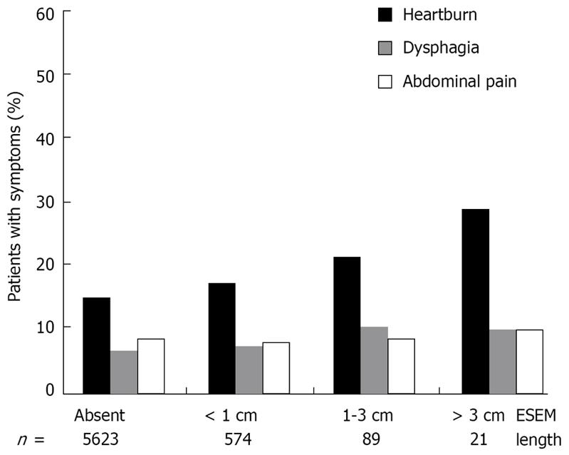
Figure 2 Correlation between ESEM length grading and each clinical symptom.
The prevalence of heartburn significantly increased in direct proportion to endoscopic ESEM length grading (P < 0.05). No significance was observed between the length of ESEM and symptoms of dysphagia and abdominal pain.
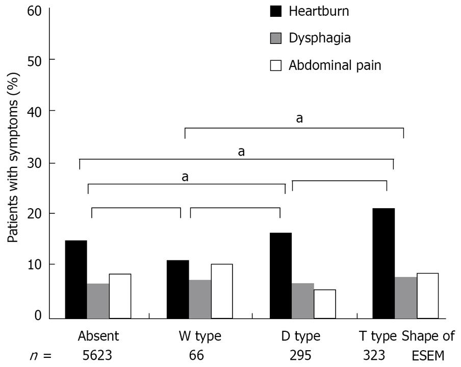
Figure 3 Correlation between the ESEM shape and each clinical symptom.
The prevalence of heartburn significantly increased in the T type ESEM than the W type ESEM and ESEM-free patients (χ2 test; aP < 0.05).
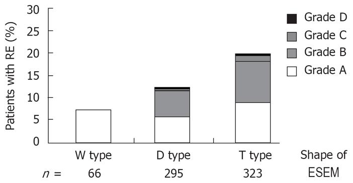
Figure 4 Correlation between the shape of ESEM and the prevalence of RE.
The prevalence of RE is significantly higher among those suffering from the T type ESEM (Kruskal-Wallis rank test, P < 0.05).
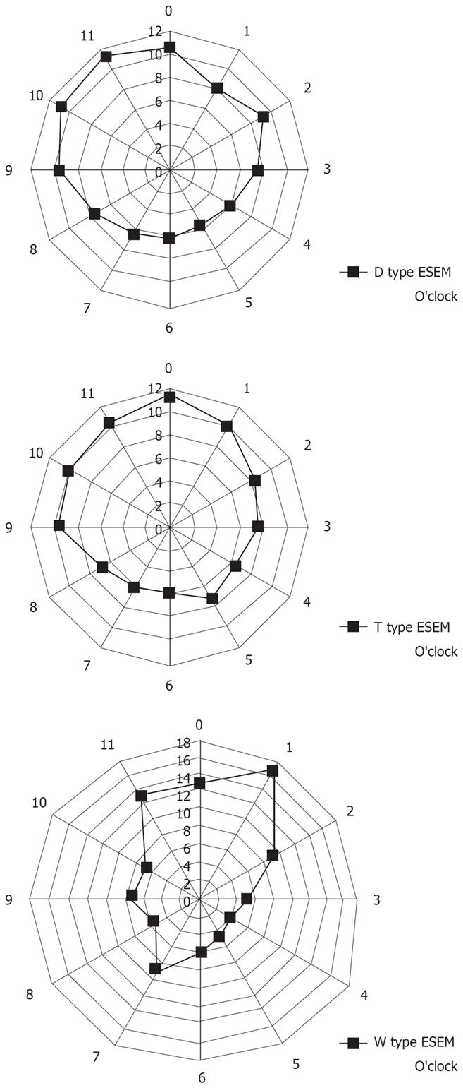
Figure 5 The circumferential location of ESEM in subjects with the W, D, and T types of ESEM.
Data are shown in terms of clock face orientation. The numbers represent the percentage of lesions on each side of the esophageal wall relative to the total number of lesions. The T-type ESEM is located mainly at the 12 and 1 o’clock wall (right anterior) of the lower esophagus, whereas the D and W types of ESEM are mainly located at the 9 to 12 o’clock wall (left anterior) of the lower esophagus. The circumferential distribution of ESEM differed significantly among subjects with different types of ESEM (χ2 test: P < 0.05, T vs D, D vs W and W vs T).
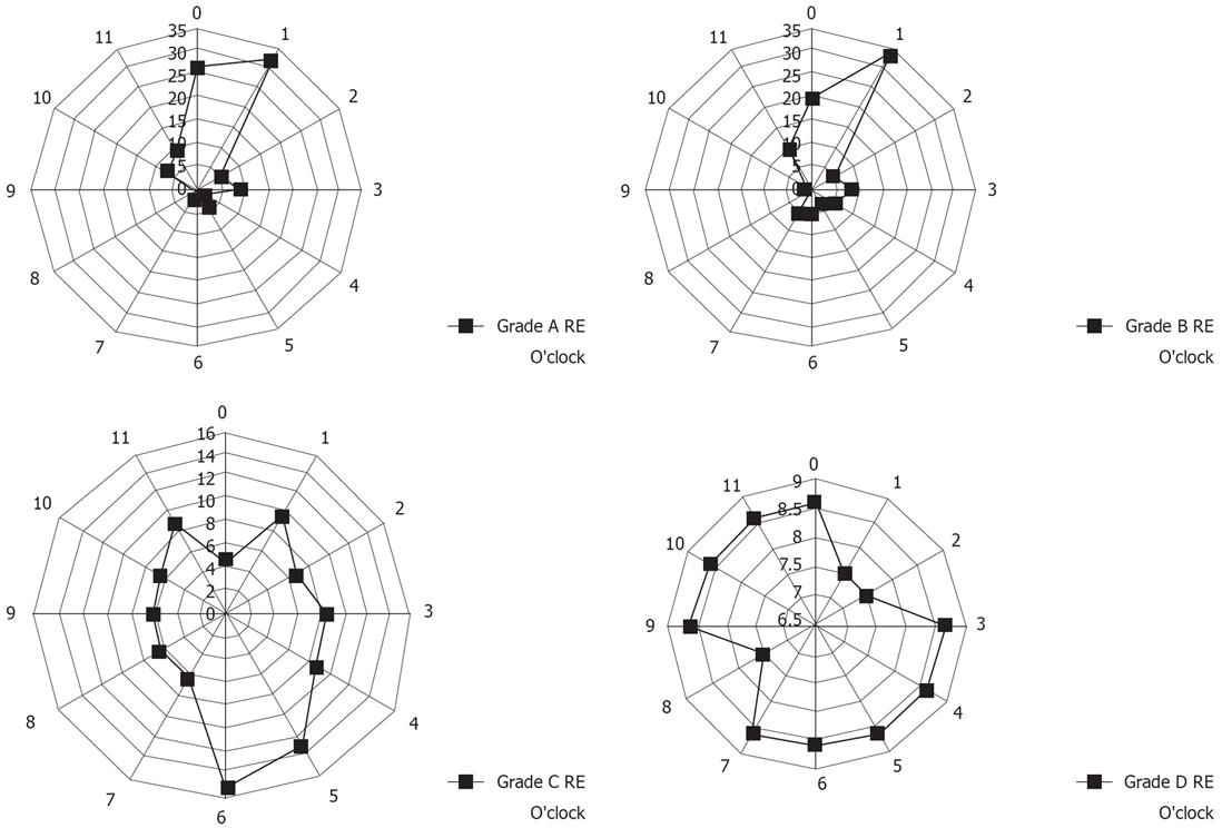
Figure 6 The circumferential location of esophageal mucosal breaks in subjects with grade A-D RE.
Data are shown in terms of clock face orientation. The numbers represent the percentage of lesions on each side of the esophageal wall relative to the total number of lesions. Patients with grade A and B esophagitis had longitudinal mucosal breaks mainly at the 12 o’clock and 1 o’clock wall of the lower esophagus, whereas patients with grade C and D esophagitis had transverse mucosal breaks mainly at the 4 o’clock to 6 o’clock wall of the lower esophagus. The circumferential distribution of esophageal mucosal breaks differed significantly among subjects with different grades of RE (χ2 test: P < 0.05, A vs B, A vs C, A vs D, B vs C, B vs D and C vs D).
- Citation: Yamagishi H, Koike T, Ohara S, Kobayashi S, Ariizumi K, Abe Y, Iijima K, Imatani A, Inomata Y, Kato K, Shibuya D, Aida S, Shimosegawa T. Tongue-like Barrett’s esophagus is associated with gastroesophageal reflux disease. World J Gastroenterol 2008; 14(26): 4196-4203
- URL: https://www.wjgnet.com/1007-9327/full/v14/i26/4196.htm
- DOI: https://dx.doi.org/10.3748/wjg.14.4196


